Dual Marker Test: Understanding Chromosomal Screening and Ayurvedic Support for Healthy Conception

ABSTRACT

Pregnancy is a remarkable phase of life in which a new individual develops from the union of two reproductive cells. During the formation of these cells and the early stages of fetal development, changes in chromosome number can occur. These chromosomal abnormalities include conditions such as Down syndrome (Trisomy 21), Edwards syndrome (Trisomy 18) and Patau syndrome (Trisomy 13). Such conditions can influence fetal growth, organ development and the health of the newborn. Modern antenatal care provides several screening procedures that help assess the possibility of chromosomal abnormalities during pregnancy. One of the important first-trimester screening procedures is the Dual Marker Test, which evaluates specific biochemical markers in maternal blood and contributes to an overall risk assessment when combined with ultrasound findings and maternal factors. At the same time, Ayurveda gives considerable importance to preconception care (Garbhadhana), reproductive health and preparation of both parents before conception. Proper nutrition, healthy lifestyle, good digestion and appropriate Ayurvedic herbs can form an important part of this preparatory phase. In this article, we shall discuss the Dual Marker Test in detail, its purpose, timing, components and interpretation, followed by a discussion of Ayurvedic preconception care and herbs that support healthy reproductive function and preparation for conception.

 Dual Marker Test

WHAT IS THE DUAL MARKER TEST?

The Dual Marker Test is a first-trimester blood screening test performed during pregnancy to assess the likelihood of certain chromosomal abnormalities. It measures two biochemical markers in the mother’s blood:

  • Free beta-human chorionic gonadotropin (free beta-hCG)
  • Pregnancy-associated plasma protein-A (PAPP-A)

These results are assessed along with factors such as maternal age, gestational age and ultrasound findings, particularly nuchal translucency (NT). The combined first-trimester screening assessment is generally performed during the early weeks of pregnancy, commonly around 11 to 14 weeks.  The purpose is not simply to produce two laboratory values. The measurements are incorporated into a calculated risk assessment that helps the obstetrician decide whether additional evaluation is appropriate.

WHY IS THE DUAL MARKER TEST IMPORTANT?

Chromosomes contain the genetic information required for growth and development.

Most human cells contain 46 chromosomes arranged into 23 pairs. During the formation of eggs and sperm, chromosomes are distributed through a specialized cell division process. An error in this process can result in an embryo receiving an additional chromosome or, in some cases, a missing chromosome.

An additional chromosome is called a trisomy. The most frequently discussed trisomies in first-trimester screening are:

Trisomy 21 — Down Syndrome

Down syndrome occurs when there is an additional copy of chromosome 21. It can be associated with characteristic physical features, developmental differences and varying degrees of intellectual disability.

Trisomy 18 — Edwards Syndrome

Edwards syndrome occurs because of an additional copy of chromosome 18. It is associated with multiple developmental and structural abnormalities.

Trisomy 13 — Patau Syndrome

Patau syndrome results from an additional copy of chromosome 13 and can affect the development of several organ systems. The Dual Marker Test forms part of screening for these chromosomal conditions.

WHAT DOES “DUAL MARKER” MEAN?

The word “dual” refers to the two biochemical markers assessed in maternal blood.

These are Free beta-hCG and PAPP-A

Both substances are associated with pregnancy and placental function. Their levels change throughout pregnancy. Therefore, the laboratory does not simply compare the measured value with one fixed reference range. The result is adjusted according to gestational age and other relevant maternal factors.

The final assessment is then combined with ultrasound findings and other information to calculate the estimated likelihood of specific chromosomal abnormalities.

FREE BETA-HCG IN THE DUAL MARKER TEST

Human chorionic gonadotropin (hCG) is an important pregnancy hormone. The free beta subunit of hCG is measured as one component of the Dual Marker Test. The concentration of free beta-hCG differs according to the stage of pregnancy. Therefore, the laboratory interprets the result according to gestational age. A value that appears high or low on its own does not provide a diagnosis. The result becomes meaningful when incorporated into the complete first-trimester screening calculation.

PAPP-A IN THE DUAL MARKER TEST

Pregnancy-associated plasma protein-A (PAPP-A) is another important marker measured during first-trimester screening. It is produced largely by placental tissue and changes during pregnancy. Like free beta-hCG, PAPP-A is interpreted according to gestational age rather than through an isolated numerical value. The combination of PAPP-A, free beta-hCG, ultrasound measurements and maternal information contributes to the final screening assessment.

WHAT IS MOM IN THE DUAL MARKER REPORT?

A Dual Marker report often includes a term called MoM, meaning Multiple of the Median. MoM compares the mother’s measured marker level with the median expected value for a particular stage of pregnancy. This allows laboratory results to be adjusted for gestational age and other variables. MoM values are therefore more useful than simply looking at the raw concentration of PAPP-A or free beta-hCG. The final risk calculation uses these adjusted values together with other pregnancy information.

ROLE OF THE NT SCAN ALONGSIDE THE DUAL MARKER TEST

The Dual Marker Test is commonly considered together with the Nuchal Translucency (NT) scan. Nuchal translucency is the measurement of fluid collected at the back of the fetal neck during an early pregnancy ultrasound. The NT measurement is obtained during a specific period of fetal development and contributes to first-trimester screening for chromosomal abnormalities. An increased NT measurement does not automatically indicate a chromosomal abnormality. It is one component of the complete assessment. The obstetrician therefore considers:

  • Maternal age
  • Gestational age
  • Free beta-hCG
  • PAPP-A
  • Nuchal translucency
  • Fetal measurements
  • Pregnancy characteristics

Before determining the overall screening result.

WHEN IS THE DUAL MARKER TEST PERFORMED?

The test belongs primarily to first-trimester screening. The combined screening assessment is generally performed around 11–14 weeks of pregnancy, depending on the pregnancy dating, fetal measurements and screening protocol. Timely pregnancy registration and regular antenatal visits help ensure that appropriate screening is performed within the recommended period. If first-trimester screening is not performed, other screening options can be discussed according to the stage of pregnancy.

HOW IS THE DUAL MARKER TEST DONE?

The blood component of the test is simple. A sample of maternal blood is collected and sent to the laboratory.

The laboratory measures Free beta-hCG and PAPP-A.

The results are then processed along with relevant maternal and fetal information. Ultrasound findings, particularly NT measurement, can be incorporated into the overall first-trimester screening assessment. The final report provides a calculated likelihood rather than confirming whether a chromosomal condition is definitely present.

SCREENING TEST VERSUS DIAGNOSTIC TEST

One of the most important things to understand about the Dual Marker Test is that it is a screening test. It does not confirm that a fetus has Down syndrome, Edwards syndrome or Patau syndrome. A higher-chance result indicates that additional assessment should be considered. A lower-chance result indicates a lower estimated likelihood but does not exclude every possible genetic or developmental condition. When additional assessment is required, the obstetrician can discuss options such as Non-Invasive Prenatal Testing (NIPT) or diagnostic procedures including Chorionic Villus Sampling (CVS) and amniocentesis.

HIGH-RISK DUAL MARKER RESULT

A higher-risk screening report can understandably create concern for parents. However, the result should always be interpreted in its proper context. A screening result represents a probability, not a confirmed diagnosis. The reported risk can be influenced by several factors, including:

  • Maternal age
  • Gestational age
  • Biochemical marker levels
  • NT measurement
  • Fetal measurements
  • Pregnancy characteristics

Therefore, a higher-risk report should lead to appropriate medical consultation rather than immediate conclusions. Additional screening or diagnostic testing can provide further information when indicated.

WHY PRECONCEPTION CARE MATTERS IN AYURVEDA

While the Dual Marker Test belongs to antenatal screening, Ayurveda places considerable importance on the period before conception. This is described through Garbhadhana (preconception care). Ayurveda gives importance to the health and nourishment of both prospective parents before conception. The aim is to prepare the body, reproductive tissues, digestion, nutrition and mental wellbeing for conception.

Important concepts include:

  • Beeja (reproductive seed)
  • Kshetra (reproductive field)
  • Ambu (nourishment)
  • Ritu (appropriate reproductive timing)
  • Shukra (male reproductive tissue)
  • Artava (female reproductive function)

This makes preconception care an important part of Ayurvedic reproductive wellness.

THE AYURVEDIC CONCEPT OF BEEJA

Beeja refers to the reproductive seed involved in conception. In modern biological terms, the egg and sperm carry genetic material contributed by the mother and father. Ayurveda places emphasis on maintaining the health and nourishment of reproductive tissues before conception. A comprehensive preconception plan therefore include:

  • Proper nutrition
  • Healthy digestion
  • Adequate sleep
  • Appropriate physical activity
  • Stress management
  • Menstrual health
  • Reproductive wellness
  • Correction of nutritional deficiencies
  • Individualized Ayurvedic herbal care

This preparatory approach provides a foundation for healthy conception.

SHATAVARI FOR PRECONCEPTION REPRODUCTIVE CARE

Shatavari (Asparagus racemosus) is one of the important Ayurvedic herbs used in women’s reproductive wellness. Its roots contain steroidal saponins, including shatavarins, along with other phytoconstituents. From an Ayurvedic perspective, Shatavari is associated with Rasayana (rejuvenative support), female reproductive nourishment and Stree Prasuti-related care. It is commonly known for:

  • Female reproductive nourishment
  • Artava support
  • General strength
  • Healthy reproductive function
  • Preconception wellness
  • Maternal nutritional support

Shatavari can be selected during the preconception period according to the woman’s constitution, digestive status, reproductive health and individual requirements. It forms an important part of Ayurvedic reproductive wellness when prescribed appropriately.

PUTRAJEEVA FOR PRECONCEPTION SUPPORT

Putrajeeva (Putranjiva roxburghii) is an Ayurvedic botanical associated with reproductive care. Its use is particularly discussed in the context of preconception wellness and reproductive preparation. Ayurvedic fertility care focuses on supporting healthy reproductive function rather than reducing the entire process of conception to a single herb. Putrajeeva can therefore be considered as part of a broader preconception regimen based on:

  • Reproductive health
  • Proper nutrition
  • Healthy digestion
  • Appropriate lifestyle
  • Individual constitution
  • Male and female reproductive factors

Its selection and formulation should be individualized by a qualified Ayurvedic physician.

SHIVLINGI FOR PRECONCEPTION REPRODUCTIVE WELLNESS

Shivlingi (Bryonia laciniosa) is another herb frequently included in Ayurvedic reproductive formulations. It is associated with Beeja-related reproductive care and is used in certain preconception approaches. Shivlingi is generally discussed as part of a broader reproductive wellness plan rather than as an isolated solution. Ayurvedic assessment can consider factors such as:

  • Prakriti
  • Agni
  • Artava
  • Shukra
  • Age
  • Reproductive history
  • Diet and lifestyle

This individualized approach helps determine whether a particular herbal formulation is appropriate for the person seeking preconception support.

SHATAVARI, PUTRAJEEVA AND SHIVLINGI: THE AYURVEDIC HERBS

These three herbs are used in Ayurvedic reproductive care because each has a place within preconception wellness. Shatavari (Asparagus racemosus) supports female reproductive nourishment and general strength. Putrajeeva (Putranjiva roxburghii) is included in selected reproductive formulations for preconception care. Shivlingi (Bryonia laciniosa) is used in certain Ayurvedic formulations associated with reproductive wellness and Beeja support. They are best considered as components of an individualized Ayurvedic program rather than as isolated remedies. The broader preconception plan should also address diet, digestion, sleep, physical activity and the reproductive health of both partners.

AYURVEDA AND MODERN PRENATAL SCREENING

Ayurvedic care and antenatal screening can be incorporated into the same pregnancy journey. The Dual Marker Test provides a first-trimester screening assessment for chromosomal conditions. Ultrasound provides information regarding fetal development and anatomy. Ayurvedic care focuses on:

  • Maternal nourishment
  • Digestive health
  • Healthy daily routine
  • Mental wellbeing
  • Appropriate lifestyle
  • Preconception reproductive health

Each approach has its own purpose. The result is a more comprehensive model of care in which reproductive wellness and appropriate antenatal screening are both given attention.

NUTRITION BEFORE CONCEPTION

Preconception nutrition deserves special attention. The body requires adequate, protein, iron, folate, vitamin B12, vitamin D, iodine, calcium, essential fatty acids, high-quality carbohydrates and dietary fiber. Adequate folate intake before conception and during early pregnancy is particularly important because it reduces the risk of neural tube defects. Ayurvedic dietary planning can be individualized according to Agni (digestive power), Prakriti (body composition), season and nutritional requirements. Food should be nourishing, digestible and appropriate for the individual’s constitution.

PREPARING BOTH PARTNERS FOR CONCEPTION

Preconception care should not focus exclusively on the woman. The health of both prospective parents contributes to conception. For women, attention should be given to:

  • Menstrual regularity
  • Nutrition
  • Reproductive health
  • Iron and folate status
  • Digestive health
  • Sleep
  • Stress management

For men, attention should be given to:

  • Shukra Dhatu (reproductive tissue)
  • Nutrition
  • Physical fitness
  • Sleep
  • Stress management
  • Avoidance of tobacco and excessive alcohol
  • General metabolic health

Ayurvedic assessment can help personalize dietary and herbal recommendations for both partners.

THE ROLE OF AGNI IN PRECONCEPTION AYURVEDA

Agni (digestive and metabolic function) occupies an important place in Ayurvedic care. Proper digestion supports the assimilation of nutrients required for tissue nourishment. A preconception program therefore focuses not only on what a person eats but also on how well the body digests and utilizes food. Appropriate meal timing, suitable food combinations, adequate sleep and individualized dietary planning support healthy Agni (digestion). When required, Ayurvedic physicians can select herbs and formulations according to the individual’s digestive status and constitution.

HEALTHY PREGNANCY BEGINS WITH RESPONSIBLE PREPARATION

A healthy pregnancy involves more than one test or one supplement.

It begins with preparation.

Before conception:

Healthy nutrition
↓
Good digestion
↓
Reproductive wellness
↓
Healthy lifestyle
↓
Individualized Ayurvedic care
↓
Preparation for conception

During pregnancy:

Regular antenatal care
↓
Appropriate ultrasound
↓
Recommended screening
↓
Maternal nutritional support
↓
Monitoring of fetal growth and development

The Dual Marker Test fits into this larger pregnancy-care framework.

CONCLUSION

The Dual Marker Test is an important first-trimester screening procedure that helps assess the likelihood of selected chromosomal abnormalities. By evaluating free beta-hCG and PAPP-A alongside ultrasound findings such as nuchal translucency and relevant maternal factors, it provides an individualized screening assessment during early pregnancy.  At the same time, Ayurveda gives special importance to the period before conception through Garbhadhana (preconception care). Attention to Beeja, Artava, Shukra, Agni, nutrition, lifestyle and mental wellbeing creates a comprehensive approach to reproductive preparation. Ayurvedic herbs such as Shatavari (Asparagus racemosus), Putrajeeva (Putranjiva roxburghii) and Shivlingi (Bryonia laciniosa) are important components of selected preconception reproductive formulations. Their role is to support reproductive wellness and preparation for conception rather than to replace genetic screening. A thoughtful approach therefore brings together preconception Ayurvedic care, nourishing nutrition, healthy lifestyle practices and appropriate antenatal screening. The Dual Marker Test provides valuable information during early pregnancy, while Ayurvedic reproductive care can begin well before conception, helping couples approach parenthood with proper preparation and comprehensive care.
Herbal Remedies of Diabetic Retinopathy – Causes, Symptoms, Diagnosis & Treatment

ABSTRACT

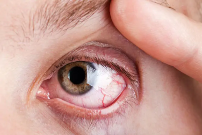
Diabetic retinopathy is a gradual eye complication associated with diabetes, in which prolonged blood sugar imbalance affects the tiny vessels nourishing the retina. The condition may begin without noticeable changes in eyesight, making regular eye examinations important. As vascular damage increases, fluid leakage, bleeding, poor circulation, retinal swelling, or abnormal vessel growth can develop. Involvement of the macula may interfere with sharp central vision and daily activities such as reading. Disease severity varies among individuals and is influenced by factors including diabetes duration, glucose levels, blood pressure, and cholesterol. Early detection, metabolic control, healthy habits, and appropriate eye care can help protect vision. 

 Diabetic Retinopathy

INTRODUCTION TO PSORIATIC ARTHRITIS

Diabetic retinopathy is a gradual eye complication associated with diabetes, in which prolonged blood sugar imbalance affects the tiny vessels nourishing the retina. The condition may begin without noticeable changes in eyesight, making regular eye examinations important.Disease severity varies among individuals and is influenced by factors including diabetes duration, glucose levels, blood pressure, and cholesterol. Early detection, metabolic control, healthy habits, and appropriate eye care can help protect vision.Our eyes depend on a healthy blood supply to work properly. At the back of the eye lies the retina, a delicate layer that receives light and sends visual information to the brain. Diabetes can gradually disturb the small blood vessels that support this important tissue. In the beginning, the person may not feel pain or notice any visual problem. With time, weakened vessels can allow fluid or blood to escape, while blocked areas may receive less oxygen. In advanced cases, fragile new vessels may appear and create further complications. This makes diabetic retinopathy an important part of long-term diabetes care, even when eyesight seems normal.

WHY DOES DIABETES AFFECT THE EYES?

Blood carries glucose throughout the body. When glucose levels remain elevated for prolonged periods, tissues exposed to this environment can undergo gradual injury.

The tiny retinal vessels are particularly sensitive. Their walls may become weaker and their ability to maintain normal blood flow can be affected. Some vessels may leak, while others may become obstructed.

Imagine a large network of very small water pipes. If some sections become weak, water may leak out. If other sections become blocked, areas beyond the blockage may receive less supply. A similar principle can help explain what happens to retinal circulation in diabetic retinopathy.Persistent vascular stress can therefore produce:

  • Leakage of fluid
  • Small retinal hemorrhages
  • Reduced blood supply
  • Retinal swelling
  • Abnormal vessel formation
  • Scar formation in advanced disease

MAJOR RISK FACTORS OF DIABETIC RETINOPATHY

The risk of diabetic retinopathy increases when diabetes and other vascular stresses remain uncontrolled for a long period. Major contributing factors include:

  • Long Duration of Diabetes – The longer a person has diabetes, the greater the chance of retinal changes developing.
  • Poor Blood Glucose Control – Persistently elevated blood sugar can gradually damage the tiny vessels supplying the retina.
  • High Blood Pressure – Hypertension places additional strain on already vulnerable blood vessels and may accelerate retinal damage.
  • Abnormal Cholesterol Levels – Disturbed lipid levels can contribute to vascular problems and are associated with diabetic eye complications.
  • Pregnancy with Pre-existing Diabetes – Pregnancy can influence the progression of existing diabetic retinopathy, making appropriate eye monitoring important.
  • Diabetic Kidney Disease – Kidney involvement can occur alongside other small-vessel complications and is associated with a greater risk of retinal disease.
  • Smoking – Tobacco use can adversely affect blood-vessel health and overall circulation.
  • Delayed Eye Screening – Because early retinopathy may have no symptoms, skipping regular retinal examinations can allow unnoticed changes to progress.
  • Poor Overall Metabolic Control – A combination of uncontrolled glucose, blood pressure, lipids and other metabolic factors can increase vascular stress.
  • Previous Retinal Changes – People who already have diabetic retinal abnormalities require appropriate follow-up because existing disease can progress.

STAGES OF DIABETIC RETINOPATHY

Diabetic retinopathy is commonly described in two broad forms: Non-proliferative diabetic retinopathy (NPDR) and proliferative diabetic retinopathy (PDR).

Non-Proliferative Diabetic Retinopathy

This is the earlier form. During this stage, the existing retinal blood vessels become abnormal. Small areas of vessel-wall weakness may appear, and minor leakage or bleeding can occur.NPDR can range from mild to severe.

  • Mild NPDR– Small vessel abnormalities may be visible during an eye examination.A person may have no noticeable change in eyesight.
  • Moderate NPDR– More retinal vessels may become affected. Leakage and areas of reduced circulation become more evident.
  • Severe NPDR- A larger portion of the retinal circulation may become compromised. The retina can experience significant oxygen shortage, increasing the possibility of abnormal vessel growth.

Proliferative Diabetic Retinopathy (PDR)

is an advanced stage of diabetic retinopathy that develops when damaged retinal blood vessels cannot provide enough oxygen to the retina. In response, the eye may form abnormal new blood vessels (neovascularization). These fragile vessels can bleed into the vitreous, causing vitreous hemorrhage, and may lead to scar tissue formation or retinal detachment. Symptoms can include blurred or suddenly reduced vision, a sudden increase in floaters or dark spots, flashes of light, difficulty seeing clearly at night, and dark areas or shadows in the field of vision. In some cases, vision may remain normal until significant bleeding or retinal damage occurs, which is why regular dilated eye examinations are important for people with diabetes.

SYMPTOMS OF DIABETIC RETINOPATHY

Diabetic retinopathy can develop gradually, and many people have no symptoms in the early stages. As retinal damage progresses, the following symptoms may appear:

  • Blurred Vision Vision may become unclear or hazy. Blurring can occur when damaged retinal blood vessels leak fluid or when swelling affects the macula, the area responsible for sharp central vision.
  • Fluctuating Vision A person may notice that eyesight changes from one day to another. Variations in blood glucose levels can temporarily affect the eye’s focusing ability, while retinal changes may also contribute to unstable vision.
  • Floaters and Dark Spots Small spots, dots, lines, or cobweb-like shapes may appear in the field of vision. A sudden increase in these floaters can occur when abnormal blood vessels bleed into the vitreous gel inside the eye.
  • Difficulty Seeing at Night Some individuals may experience reduced ability to see clearly in dim light or may find nighttime activities more difficult as retinal function becomes affected.
  • Dark or Empty Areas in Vision Damaged or poorly supplied portions of the retina can cause missing areas in the visual field. These may appear as patches of darkness or areas where objects are difficult to see.
  • Reduced Central Vision When diabetic macular edema affects the macula, detailed central vision may become impaired. This can make reading, writing, recognizing faces, or seeing fine details more difficult.
  • Distorted Vision Straight lines may appear bent, wavy, or irregular when swelling or structural changes involve the macula.
  • Changes in Color Vision Colors may appear less bright or more difficult to distinguish. Retinal damage can interfere with the normal processing of visual information.
  • Sudden Vision Changes A rapid decrease in eyesight may occur because of bleeding from fragile abnormal blood vessels, particularly in proliferative diabetic retinopathy.
  • Partial or Severe Vision Loss Advanced diabetic retinopathy can cause significant loss of sight. Complications such as vitreous hemorrhage or retinal detachment can result in serious visual impairment.
  • Flashes of Light Flashes may occur when abnormal pulling or traction develops inside the eye, particularly when scar tissue forms around abnormal blood vessels.
  • Curtain or Shadow Over Vision A curtain-like shadow across part of the visual field can be a warning sign of retinal detachment and requires urgent ophthalmic assessment.

INVESTIGATIONS

The diagnosis and assessment of diabetic retinopathy involve a combination of eye examination, retinal imaging, and systemic investigations. Important investigations include:

Comprehensive Dilated Eye Examination

  • The pupils are dilated so the ophthalmologist can examine the retina, macula, optic nerve, and retinal blood vessels.
  • It helps identify microaneurysms, retinal hemorrhages, exudates, macular edema, and abnormal new blood vessels.

Visual Acuity Test

  • Measures how clearly the patient can see at different distances.
  • It helps assess the functional effect of retinal disease on vision.

Fundus Photography

  • Photographs of the retina are taken to document retinal changes.
  • Serial photographs can help monitor progression over time.

Optical Coherence Tomography (OCT)

  • OCT provides detailed cross-sectional images of the retina.
  • It is particularly useful for detecting diabetic macular edema (DME) and measuring retinal thickening.

Optical Coherence Tomography Angiography (OCTA)

  • Provides images of retinal and choroidal blood-flow patterns without the need for injected dye.
  • It can help identify areas of reduced retinal perfusion and abnormal vascular changes.

Fluorescein Angiography (FFA)

  • A fluorescent dye is injected into a vein and photographs are taken as the dye circulates through retinal vessels.
  • It can demonstrate leakage, blocked areas of circulation, and abnormal new blood vessels and can help guide laser treatment.

B-Scan Ultrasonography

  • Useful when the retina cannot be clearly visualized because of vitreous hemorrhage or other media opacity.
  • It can help assess conditions such as retinal detachment.

Systemic Investigations

  • HbA1c and blood glucose: assess long-term and current glycemic control.
  • Blood pressure: hypertension can increase the risk of retinal complications.
  • Lipid profile: helps identify associated cardiovascular risk factors.
  • Kidney function tests: may be appropriate because diabetic kidney disease and retinopathy can occur together.

TREATMENT OF DIABETIC RETINOPATHY

stage of retinopathy, presence of macular edema, visual symptoms, and overall health. Some patients require monitoring alone, while advanced disease may require active treatment.

Optimizing Diabetes and Cardiovascular Risk Factors

Good control of blood glucose, blood pressure, and cholesterol is an important part of diabetic retinopathy management. Smoking cessation and appropriate management of associated medical conditions are also important.

This approach does not replace eye-specific treatment when advanced retinal disease is present, but it helps reduce the risk of progression.

Anti-VEGF Injections

Anti-vascular endothelial growth factor (anti-VEGF) medicines are commonly used for diabetic macular edema and certain cases of proliferative diabetic retinopathy.They work by reducing the activity of VEGF, a signaling protein involved in abnormal blood-vessel growth and vascular leakage.

Treatment generally involves injections into the vitreous of the eye, with the number and frequency determined by the ophthalmologist.

Laser Photocoagulation

Laser treatment may be used in selected cases, particularly for proliferative diabetic retinopathy and certain forms of diabetic macular edema.

In proliferative disease, panretinal photocoagulation (PRP) reduces the oxygen demand of peripheral retinal tissue and can help reduce the stimulus for abnormal blood-vessel growth.

Intravitreal Corticosteroids

Corticosteroids may be considered for selected patients with diabetic macular edema, particularly when anti-VEGF therapy is unsuitable, insufficient, or when the ophthalmologist considers steroids appropriate.

Because corticosteroids can increase the risk of cataract and elevated intraocular pressure, patients receiving them require appropriate monitoring.

Vitrectomy Surgery

A vitrectomy may be required in advanced complications such as:

  • Persistent or non-clearing vitreous hemorrhage
  • Tractional retinal detachment involving or threatening the macula
  • Certain severe proliferative complications
  • Other situations where the vitreous is preventing appropriate retinal management

AYURVEDA PERSPECTIVE OF DIABETIC RETINOPATHY

Diabetic Retinopathy can be understood from an Ayurvedic perspective through the concepts of Prameha (urinary-metabolic disorders), Madhumeha (a Vata-dominant form of Prameha), Timira (visual disturbance), Dosha-Dushti (functional imbalance of Doshas), Dhatu-Dushti (tissue disturbance), Rakta Dushti (vitiation of blood tissue), Srotodushti (disturbance of body channels), Agnimandya (weak digestive and metabolic activity) and Ama (incompletely processed metabolic material). Ayurveda does not describe diabetic retinopathy as an exact single disease entity in the classical texts. Therefore, its Ayurvedic understanding is a conceptual correlation, rather than a direct classical equivalence.

The progression can be viewed as a long-standing disturbance beginning with Agni, followed by improper processing of food and tissues, Dosha aggravation and disturbance of Dhatus and Srotas. In Madhumeha, prolonged involvement of Kapha, Pitta and Vata, together with Meda (fat tissue), Kleda (excess fluid/metabolic moisture), Rakta (blood tissue) and other Dhatus, may gradually affect the delicate structures responsible for vision. The eye, particularly Drishti (visual apparatus/vision) and its Patala (layers or anatomical coverings), becomes involved when the disturbed Doshas reach the ocular tissues. Classical Ayurvedic descriptions of Timira explain how aggravated Doshas affecting the ocular Patala can produce blurred or dim vision.

Thus, Madhumehajanya Timira may be used as an Ayurvedic framework for understanding diabetic retinal changes, with emphasis on maintaining Agni, controlling Prameha, balancing Doshas, protecting Dhatus, maintaining healthy Srotas and supporting Chakshus (eyes/vision).

Nidana (Causative Factors)

The causative factors can be considered at two levels:

Factors contributing to Prameha

Ayurveda places strong emphasis on inappropriate Ahara (diet) and Vihara (lifestyle).
Important factors may include:

  • Excessive Madhura Ahara (sweet foods)
  • Excessive Guru Ahara (heavy foods)
  • Excessive Snigdha Ahara (unctuous foods)
  • Excessive food intake
  • Lack of physical activity
  • Excessive daytime sleeping
  • Sedentary habits
  • Repeated overeating
  • Improper daily routine
  • Excessive Kapha-promoting dietary habits

These factors may contribute to Kapha, Meda and Kleda Dushti, followed by disturbance of Agni and Prameha.

Factors contributing to ocular involvement

When Prameha becomes chronic, continued Dosha and Dhatu disturbance may create a background for ocular involvement.
The conceptual pathway is:-Prameha → Madhumeha → chronic Dosha-Dhatu Dushti → Rakta/Srotas involvement → Netra Dushti → Drishti Vikara

SAMPRAPTI (DISEASE-DEVELOPMENT PROCESS)

Mithya Ahara-Vihara (inappropriate diet and lifestyle)
↓
Agnimandya (weak digestive and metabolic activity)
↓
Ama formation (improperly processed metabolic material)
↓
Dosha Prakopa (aggravation of Doshas)
↓
Meda and Kleda Dushti (disturbance of fat tissue and fluid components)
↓
Prameha/Madhumeha
↓
Dhatu Dushti (tissue disturbance)
↓
Rakta Dushti (blood-tissue disturbance)
↓
Srotodushti (channel disturbance)
↓
Netra involvement (ocular involvement)
↓
Drishti Dushti (visual disturbance)
↓
Madhumehajanya Timira.

SYMPTOMS

From an Ayurvedic perspective, manifestations of Madhumehajanya Timira may be discussed under Drishti Vikara (visual disturbance).Possible features include:

Timira (dimness of vision)

The person may feel that vision has become less clear or objects appear hazy.

Avyakta Darshana (unclear vision)

Objects may not appear sharply defined, particularly when the ocular disturbance progresses.

Drishti Mandya (reduced visual ability)

There may be a gradual decline in the ability to perceive objects clearly.

Netra Gaurava (heaviness of the eyes)

The eyes may feel tired or heavy, particularly in individuals with associated Dosha imbalance.

Daha (burning sensation)

Pitta involvement may be considered when burning or heat-related discomfort is present.

Raga (redness)

Rakta-Pitta involvement may be considered when redness accompanies ocular symptoms.

Ruja (ocular discomfort or pain)

Pain is not necessarily present in every case and depends on the underlying ocular condition.

Drishti Bhrama (disturbed visual perception)

The person may experience altered or uncomfortable visual perception.

Progressive Drishti Hani (decline in vision)

With increasing ocular involvement, visual function may gradually become impaired.

MANAGEMENT OF MADHUMEHA/PRAMEHA

Protection and support of Netra and Drishti. The major Ayurvedic principles include:

Agni Deepana (supporting digestive and metabolic fire)

Improving Agni is considered important when Agnimandya is present.
The objective is to support proper digestion and metabolism and reduce the tendency toward Ama formation.

Ama Pachana (metabolic cleansing)

Where Ama is considered present, appropriate Ama Pachana measures may be used according to the individual’s condition.

Prameha Chikitsa (management of Prameha)

The underlying metabolic disorder should be addressed through appropriate Ahara, Vihara and classical Ayurvedic management.

Dosha Shamana (Dosha balancing)

The predominant Dosha should be assessed before selecting treatment.

Meda-Kleda Management

Since Meda and Kleda are important in the Ayurvedic pathogenesis of Prameha, appropriate measures are selected to maintain their balance.

Rakta Prasadana (supporting healthy Rakta)

Where Rakta Dushti is considered, appropriate Ayurvedic measures may be selected according to the patient’s Dosha and clinical presentation.

Srotoshodhana (supporting healthy channel function)

Management aims to reduce factors contributing to Srotorodha (channel obstruction) and maintain normal movement and nourishment.

Rasayana (rejuvenative support)

Rasayana is used in Ayurveda to support tissue strength, nourishment and resilience.
In chronic disorders, Rasayana may be considered according to the patient’s age, strength, Agni, Dosha and disease stage.

HERBAL REMEDIES FOR DIABETIC RETINOPATHY BY PLANET AYURVEDA

Planet Ayurveda is an Ayurvedic wellness brand focused on bringing traditional Ayurvedic knowledge into everyday life . Its products are developed around the principles of Ayurveda and commonly used herbs, classical ingredients, and traditional formulations. The brand aims to make Ayurvedic care easier to understand and use by combining traditional concepts with a modern approach to health and wellness. Its range includes herbal formulations designed for different areas of wellbeing, including digestion, joints, metabolism, immunity, and general health. Planet Ayurveda emphasizes the use of naturally sourced Ayurvedic ingredients and presents them in convenient forms such as capsules, tablets, powders, and other formulations.The management of Madhumehajanya Timira can be understood through two interconnected goals

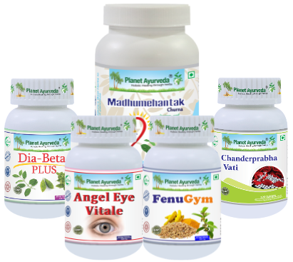
  1. Chandraprabha Vati
  2. Diabeta Plus Capsules
  3. Fenugym Capsules
  4. Madhumehantak Churna
  5. Angel Eye Vitale Capsules

Herbal Remedies For Diabetic Retinopathy

Buy Now Herbal Remedies For Diabetic Retinopathy

Buy Now Herbal Remedies For Diabetic Retinopathy

PRODUCT DIISCRIPTION

1. Chandraprabha Vati

This is a classical Ayurvedic tablet formulation traditionally used to support urinary function, digestion, metabolism, and general vitality. It contains traditionally valued ingredients such as Guduchi (Tinospora cordifolia), Guggul (Commiphora mukul), Shilajit (Asphaltum punjabianum), Haridra (Curcuma longa), Amalaki (Emblica officinalis), Shunthi (Zingiber officinale)  and mineral salts. These herbs are traditionally used for metabolic and rejuvenative support, while the Deepana-Pachana herbs support healthy digestion and Agni.

Dosage– 1–2 tablets twice daily with lukewarm water

2. Diabeta Plus Capsules

These capsules are an Ayurvedic herbal formulation traditionally used as supportive care for Madhumeha and to maintain healthy glucose metabolism. The formulation combines Karela (Momordica charantia), Vijaysar (Pterocarpus marsupium), Gurmar (Gymnema sylvestre), Saptarangi (Salacia chinensis), Ashwagandha (Withania somnifera) etc. These herbs are traditionally valued for metabolic and digestive support and commonly used in Ayurvedic practice for supporting healthy glucose and carbohydrate metabolism. Ashwagandha is traditionally regarded as a Rasayana and supports strength, vitality, and general wellbeing, while Tulsi provides traditional digestive and antioxidant support.

Dosage– 1–2 capsules twice daily with plain water.

3. Fenugym Capsules

These capsules are an Ayurvedic herbal formulation designed to provide supportive care for healthy glucose levels, digestion, and metabolic balance. It combines four traditionally used herbs: Saptarangi, Fenugreek, Haridra, and Gurmar. Saptarangi is traditionally associated with Prameha care and carbohydrate metabolism, while Fenugreek is valued for supporting digestion and healthy carbohydrate handling. Gurmar is commonly used in Ayurvedic practice for Madhumeha and metabolic support. Haridra is traditionally valued for its antioxidant and anti-inflammatory properties. Together, these herbs provide comprehensive traditional support for maintaining healthy metabolic function

Dosage–  1–2 capsules twice daily

4. Madhumehantak Churna

This is an Ayurvedic herbal powder traditionally used as supportive care for healthy blood sugar levels, digestion, and metabolic function. Its formulation includes Gudmar (Gymnema sylvestre), Methi (Trigonella foenum-graecum), Jamun Beej (Syzygium cumini), Karela (Momordica charantia), Neem (Azadirachta indica), Haridra (Curcuma longa) are traditionally associated with Madhumeha and healthy glucose metabolism, while Methi supports digestion and carbohydrate metabolism. Neem and Haridra provide traditional bitter, cleansing, antioxidant, and metabolic support. Kutki is traditionally valued for supporting liver and digestive function. This formulation is available as a churna (herbal powder).

Dosage– 1–2 teaspoons twice daily

5. Angel Eye Vitale Capsules

These are an Ayurvedic herbal formulation designed to provide traditional nutritional and rejuvenative support for healthy eyes and normal vision. The formulation contains selected herbs including Gotu Kola (Mandukaparni), Bhringraj, and Amla, traditionally valued for eye nourishment and overall wellbeing. Gotu Kola (Centella asiatica) is traditionally used to support healthy vision and nourish the eyes. Bhringraj (Eclipta alba) is traditionally regarded as a nourishing and rejuvenative herb, while Amla (Emblica officinalis) is naturally rich in antioxidants and traditionally used for eye and systemic support.

Dosage– 1–2 capsules twice daily

CONCLUSION

Diabetic retinopathy is a serious eye complication of diabetes that can gradually damage the small blood vessels of the retina. In the early stages, it may develop without clear symptoms, which makes regular eye examinations and timely screening very important. Good control of blood glucose, blood pressure, and cholesterol can help reduce the risk of progression. Treatment depends on the stage and may include medical management, laser therapy, injections, or surgery when required. From an Ayurvedic perspective, supportive measures such as a balanced Pathya Ahara (appropriate diet), healthy lifestyle, and selected herbal formulations may be used to support overall metabolic and eye health. However, Ayurvedic care should complement, rather than replace, regular ophthalmic evaluation and indicated treatment. Early detection, consistent diabetes management, and proper follow-up are key to protecting vision and reducing the risk of severe visual loss.

Herbal Remedies of Psoriatic Arthritis – Causes, Symptoms, Diagnosis & Treatment

ABSTRACT

Psoriatic arthritis (PsA) is a chronic inflammatory condition that affects the joints and may occur along with psoriasis. It develops when the immune system becomes overactive and triggers inflammation in different parts of the musculoskeletal system. Patients may experience joint pain, swelling, morning stiffness, tenderness, and difficulty moving, while some also develop swelling of entire fingers or toes and discomfort around tendons. The pattern and severity can differ greatly between individuals. If persistent inflammation remains uncontrolled, structural changes and loss of joint function may occur. Early identification and appropriate management are important for maintaining mobility and quality of life. A comprehensive approach may include medical treatment, physical activity, weight management, balanced nutrition, stress control, and supportive therapies. Understanding the connection between skin, immune activity, and joint health can help in developing a more personalized approach to long-term care.

 Psoriatic Arthritis

INTRODUCTION TO PSORIATIC ARTHRITIS

Psoriatic arthritis (PsA) is a long-term inflammatory joint condition that can develop in people with psoriasis, a skin disorder characterized by red, scaly patches. Unlike ordinary wear-and-tear arthritis, PsA involves an abnormal immune response that can affect the joints, tendons, ligaments, fingers, toes, and spine. It may cause pain, stiffness, swelling, and reduced movement, with symptoms often varying from person to person. Some individuals develop joint problems before noticeable skin changes, making early recognition important.The disease can gradually damage affected joints if persistent inflammation is not adequately controlled. Psoriatic arthritis is therefore more than a skin-related problem—it is a systemic inflammatory disorder requiring attention to both musculoskeletal and overall health.

MECHANISM OF ACTION

Psoriatic arthritis (PsA) is a chronic immune-mediated inflammatory arthritis associated with psoriasis. The process begins with abnormal activation of the immune system, particularly involving dendritic cells, T cells, and inflammatory cytokines such as TNF-α, IL-17, and IL-23. The IL-23/IL-17 pathway plays an important role in maintaining inflammation at the joints, entheses (where tendons and ligaments attach to bone), and skin. These inflammatory signals stimulate synovial cells and other tissues, leading to synovitis, swelling, pain, and stiffness. At the same time, inflammation at the entheses can cause enthesitis, while chronic inflammation can promote both bone erosion and abnormal new bone formation. This combination of tissue inflammation, bone damage, and new bone growth contributes to the characteristic joint changes and reduced mobility seen in PsA.
The mechanism can be summarized as:inflammatory cytokine → receptor activation

cytokine signal
↓
jak activation
↓
stat activation
↓
nuclear signaling
↓
inflammatory gene expression
↓
inflammatory response
with jak1 inhibition

↓
reduced jak1-mediated signaling
↓
reduced downstream cytokine signaling
↓
lower inflammatory activity.

SYMPTOMS OF PSORIATIC ARTHRITIS

JOINT PAIN AND SWELLING

Peripheral arthritis is one of the most common manifestations. Patients may notice tenderness, swelling, warmth, or difficulty moving affected joints. hands and feet may be particularly troublesome, although knees, ankles, wrists, and other joints can also become involved.The pattern may be asymmetric or affect multiple joints on both sides of the body. 

MORNING STIFFNESS

Inflammatory Stiffness is often more noticeable after prolonged rest.

ENTHESITIS

Enthesitis refers to inflammation where tendons, ligaments, or fascia attach to bone.Commonly affected areas include:

  • Achilles Tendon
  • Plantar Fascia
  • Knees
  • Elbows
  • Pelvic Attachment Sites.

DACTYLITIS

Dactylitis produces diffuse swelling of an entire finger or toe, giving the digit a characteristic enlarged appearance.It results from inflammation involving several structures simultaneously, including joints, tendon sheaths, and surrounding tissues.

AXIAL SYMPTOMS

Some individuals develop inflammation affecting the spine or sacroiliac joints.

SKIN MANIFESTATIONS

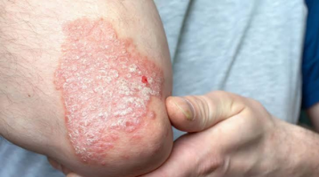
Psoriasis may appear as well-defined plaques covered with scales. The scalp, elbows, knees, trunk, and other areas can be involved.

NAIL CHANGES

  • Nail involvement may include:
  • Pitting
  • Ridging
  • Thickening
  • Discoloration
  • Separation of the nail from its bed

FATIGUE AND FUNCTIONAL DIFFICULTIES

Chronic inflammation can be associated with fatigue and reduced physical capacity. Difficulty opening containers, writing, walking, exercising, climbing stairs, or performing occupational tasks may develop when disease activity remains uncontrolled.

INVESTIGATIONS FOR PSORIATIC ARTHRITIS

There is no single blood test that independently confirms psa. diagnosis is based on the clinical picture, physical examination, medical history, psoriasis status, musculoskeletal findings, laboratory assessment, and imaging when necessary.

CLINICAL EVALUATION

  • The first investigation is a detailed clinical assessment.
  • Number And Distribution Of Tender Joints.
  • Number Of Swollen Joints.
  • Presence Of Dactylitis.
  • Enthesitis Sites.
  • Spinal Mobility.
  • Sacroiliac Symptoms.
  • Nail Abnormalities.
  • Extent Of Psoriasis.
  • Functional Limitations.
  • Previous Episodes Of Inflammatory Disease.

BLOOD INVESTIGATIONS

Blood tests in psoriatic arthritis (PsA) mainly help assess inflammation, anemia, and exclude other types of arthritis.

COMPLETE BLOOD COUNT

Hemoglobin (Hb): May be decreased, particularly with chronic inflammation, resulting in anemia of chronic disease. Total Leukocyte Count (TLC/WBC)-May be normal or mildly increased due to systemic inflammation. Platelet Count (PLT): Can be increased (thrombocytosis) during active inflammation; it may remain normal in mild or controlled disease.

ESR

Erythrocyte sedimentation rate can provide an indirect indication of systemic inflammatory activity.

CRP

C-Reactive Protein Is Another Marker Used To Assess Inflammation.

RHEUMATOID FACTOR

Rheumatoid Factor May Be Measured When Rheumatoid Arthritis Is Part Of The Differential Diagnosis. PSA  Is Commonly Seronegative For Rheumatoid Factor.

ANTI-CCP ANTIBODIES

Anti-Cyclic Citrullinated Peptide Antibodies May Also Help Distinguish Rheumatoid Arthritis From Psa In Appropriate Clinical Situations.

X-RAY

  • Conventional Radiography Can Demonstrate Structural Changes Such As:
  • Erosions
  • Joint-Space Alterations
  • New Bone Formation
  • Ankylosis
  • Other Chronic Changes

ULTRASOUND

Musculoskeletal Ultrasound Can Identify:

  • Synovitis
  • Tendon Sheath Inflammation
  • Enthesitis
  • Increased Blood Flow Associated With Active Inflammation.

DIFFERENTIAL DIAGNOSIS

Because Several Diseases Can Produce Joint Pain And Swelling, Psa Must Be Differentiated From Other Inflammatory And Musculoskeletal Disorders.Important Possibilities Include:

  • Rheumatoid Arthritis
  • Osteoarthritis
  • Gout
  • Reactive Arthritis
  • Ankylosing Spondylitis And Related Spondyloarthritis

AYURVEDIC PERSPECTIVE OF PSORIATIC ARTHRITIS

Psoriatic arthritis (psa) does not have a single, exact equivalent in the classical ayurvedic texts. Its combination of chronic skin lesions and inflammatory musculoskeletal manifestations can be understood through overlapping ayurvedic concepts, particularly kushtha (chronic skin disorders), vatarakta (vata-rakta disorder), and amavata (ama-associated joint disorder), depending upon the patient’s individual presentation. Therefore, an ayurvedic correlation should be based on the dominant dosha (functional biological principle), Dhatu (body tissue), Srotas (physiological channel), agni (digestive and metabolic capacity), and ama (incompletely processed metabolic material) rather than assigning every patient to one classical diagnosis.

The skin component may be considered in relation to Twak (skin) and Rakta Dhatu (blood tissue), while pain, stiffness, swelling, and restricted joint movement indicate involvement of sandhi (joints) and predominantly vata dosha. kapha may contribute to heaviness, swelling, itching, and thickening. Thus, the ayurvedic picture may involve a combination of vata-kapha imbalance, rakta involvement, impaired agni, and ama, with the exact pattern varying from person to person.

The ayurvedic pathogenesis may begin with Mithya ahara-vihara (improper diet and lifestyle), irregular eating, excessive heavy food, unsuitable food combinations, sedentary habits, inadequate sleep, chronic stress, or other individual triggers.

These factors may disturb agni (digestive and metabolic function).
agni dushti (impaired digestion and metabolism)
↓
ama utpatti (formation of ama)
↓
dosha prakopa (aggravation of doshas)
↓
srotodushti (disturbance of body channels)
↓
rakta and twak involvement (skin-related manifestation)
↓
vata involvement in sandhi (joint localization)
↓

sandhi shoola + shotha + stambha
(joint pain + swelling + stiffness

  • NIDANA PARIVARJANA – AVOIDANCE OF CAUSATIVE FACTORS- The first principle is to identify and reduce factors that aggravate the patient’s condition.This may include:
  • irregular meals
  • overeating
  • excessively heavy food
  • highly processed foods
  • excessive sedentary behavior
  • poor sleep routine
  • excessive stress
  • individually identified dietary triggers
  • AGNI DEEPANA – SUPPORTING DIGESTIVE FUNCTION- When impaired digestion is present, treatment may emphasize agni deepana (supporting digestive capacity).
  • AMA PACHANA –When the clinical picture indicates ama, ama pachana (supporting the processing and reduction of ama) may form part of the treatment strategy.
  • DHATU SUPPORT – TISSUE NOURISHMENT-Chronic musculoskeletal disease requires attention to asthi dhatu (bone tissue), mamsa dhatu (muscle tissue), majja dhatu (marrow/deeper tissue concept), and rakta dhatu (blood tissue) according to the patient’s presentation.

HERBAL REMEDIES FOR PSORIATIC ARTHRITIS  BY PLANET AYURVEDA

Planet Ayurveda is an Ayurvedic wellness brand focused on providing herbal formulations based on the principles of Ayurveda (traditional Indian system of medicine). The brand offers a range of products prepared using herbs, herbal extracts, classical Ayurvedic ingredients, and traditional formulations.  Planet Ayurveda emphasizes the traditional Ayurvedic concepts of Dosha balance, Agni (digestive fire), Dhatu nourishment (tissue health), and natural herbal support. Products are presented as supportive wellness options and are intended to complement healthy lifestyle practices. Individual products should be selected according to the person’s condition and used appropriately, particularly when someone has a diagnosed medical condition or is taking conventional medicines.Here, we will discuss  formulations used for psoriatic arthritis.

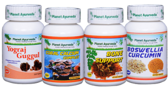
  1. Boswellia-Curcumin
  2. Yograj Guggul
  3. Coral Calcium Complex
  4. Bone Support

Herbal Remedies For Psoriatic Arthritis

Buy Now Herbal Remedies For Psoriatic Arthritis

Buy Now Herbal Remedies Psoriatic Arthritis

PRODUCT DESCRIPTION

1. Boswellia–Curcumin

Boswellia–Curcumin combines two well-known herbs, Shallaki (Boswellia serrata) and Haridra (Curcuma longa), traditionally used for Shotha (inflammation) and Sandhi Shoola (joint pain). Shallaki supports a balanced inflammatory response and may help reduce Sandhi Shotha (joint swelling), stiffness, and discomfort.its resin contains boswellic acids, which have been investigated for their effects on inflammatory pathways. Haridra is valued for its Shothahara (anti-inflammatory) and Vedanasthapana (pain-relieving) properties and provides antioxidant support. its major bioactive constituent, curcumin, has been investigated for interactions with multiple inflammatory pathways.Together, they may support comfortable joint movement and help maintain Sandhi Bala (joint strength), making them useful as supportive care for inflammatory joint conditions such as psoriaticArthritis.

Dosage -1 Capsule Twice Daily .

2. Yograj Guggulu

Yograj guggulu is a classical ayurvedic formulation prepared using Guggul (Commiphora mukul) etc traditionally used in disorders involving vata dosha and the musculoskeletal system.it is traditionally considered in presentations involving:sandhi shoola (joint pain),sandhi stambha (joint stiffness),reduced mobility,vata-associated musculoskeletal discomfort.Its role can be understood within the broader ayurvedic strategy of supporting normal vata function and musculoskeletal mobility.

Dosage -1 Tablet Twice A Day.

3. Coral Calcium Complex

Coral calcium Complex capsules is used as a nutritional component of a bone-support strategy prepared using Guduchi (Tinospora Cordifolia), Kamdudha Ras etc. In ayurvedic terminology, Asthi dhatu (bone tissue) is responsible for maintaining the skeletal framework of the body. Adequate nutritional support is important for maintaining normal bone mineralization.This combination brings together Pitta-Shamana (Pitta-balancing), Daha-Shamana (relief of burning/heat), Rasayana (rejuvenative support), and Dhatu Poshan (tissue nourishment) concepts.

Dosage – 1 Capsule Twice Daily .

4. Bone support

Bone support capsules by Planet Ayurveda are prepared using Arjuna (Terminalia arjuna), Hadjod (Cissus Quadrangularis) etc. A bone support formulation can be incorporated into an ayurvedic supportive program when maintaining skeletal health is an important objective.Ayurveda considers asthi dhatu as an important component of the musculoskeletal system,while modern medicine recognizes the importance of adequate calcium, vitamin d, protein, physical activity, and muscle strength for skeletal health.

Dosage– 1-2 Capsule Twice Daily.

DIET FOR PSORIATIC ARTHRITIS

According to Ayurveda, dietary planning in Psoriatic Arthritis can focus on balancing aggravated Vata and Pitta Dosha, reducing Ama (metabolic waste/undigested material), supporting Agni (digestive fire), and avoiding foods that may promote Shotha (inflammation).
Recommended Foods

  • Laghu Ahara (light food): Prefer easily digestible meals such as Yavagu (thin rice porridge), Mudga Yusha (moong dal soup), Khichadi, and vegetable soups.
  • Mudga (green gram): Light and relatively easy to digest; useful when Agni is weak.
  • Shali Dhanya (old rice): Traditionally considered light and suitable for regular meals.
  • Yava (barley): Can be included in moderation as a Laghu and Kapha-reducing grain.
  • Tikta Shaka (bitter vegetables): Vegetables such as Patola (pointed gourd), Karela (bitter gourd), and leafy greens may be included according to individual tolerance.
  • Drava Ahara (liquid foods): Warm soups and thin preparations can be useful when digestion is weak.
  • Ushna Jala (warm water): Prefer warm or comfortably hot water instead of frequent chilled beverages.
  • Haridra (turmeric): May be incorporated into food as a traditional Shothahara (inflammation-supporting) spice.
  • Shunthi (dry ginger): Used traditionally to support Agni Deepana (digestion stimulation) and reduce Ama.
  • Jeeraka (cumin): Can be used in cooking to support Deepana-Pachana (digestive function).
  • Dhaniya (coriander): May be used as a mild digestive spice, particularly when Pitta is prominent.

FOODS TO LIMIT

  • Guru Ahara (heavy foods): Excessive fried, greasy, and very rich foods may be difficult to digest and can contribute to Ama formation.
  • Abhishyandi Ahara (foods that promote obstruction/kapha accumulation): Excessive curd, cheese, cream, and very heavy dairy preparations should be limited, especially when there is Kapha or Ama predominance.
  • Dadhi (curd/yogurt): Ayurveda traditionally advises caution with frequent or excessive curd consumption, particularly in inflammatory and Kapha-associated conditions.
  • Madhura Ati-Sevana (excessive sweet foods): Reduce sweets, sugary beverages, desserts, and highly processed foods.
  • Ati-Lavana (excess salt): Avoid excessive salty and packaged foods.
  • Amla Ahara (excessively sour foods): Limit very sour, pickled, and fermented foods when Pitta is aggravated.
  • Katu Ahara (excessively pungent foods): Very spicy foods may aggravate Pitta in susceptible individuals.
  • Viruddha Ahara (incompatible food combinations): Avoid traditionally incompatible combinations, particularly Dadhi with certain foods and other combinations that are poorly tolerated.
  • Guru-Mamsa (heavy meats): Excessive intake of heavy, fatty meat preparations should be avoided when digestion is weak.
  • Madya (alcohol): Avoid or minimize alcohol, as it may aggravate inflammatory symptoms and is traditionally considered unsuitable in many inflammatory conditions.

CONCLUSION

Psoriatic arthritis can be interpreted as a complex disorder involving dosha imbalance, impaired agni, ama, rakta and twak involvement, and vata-related disturbance of sandhi, with the exact presentation differing between individuals. The combination of skin manifestations and musculoskeletal symptoms makes individualized assessment especially important.The ayurvedic strategy therefore extends beyond the use of a single formulation. it may involve nidana parivarjana (removal of causative factors), agni deepana (supporting digestion), ama pachana (addressing ama), dosha shamana (pacifying aggravated doshas), appropriate diet and dinacharya (daily routine), physical activity, and selected supportive formulations.Within this framework, boswellia-curcumin may be positioned for joint and inflammatory support, yograj guggulu for traditional vata-related musculoskeletal support, coral calcium for calcium supplementation and skeletal nutrition, and bone support for maintaining overall bone health.A responsible integrative approach therefore combines appropriate rheumatological disease control with individualized ayurvedic lifestyle and supportive measures, while ensuring that potential herb–drug interactions, contraindications, product quality, and ongoing disease activity are reviewed by qualified healthcare professionals.

Herbal Remedies of Mother Fixation – Causes, Symptoms, Diagnosis & Treatment

ABSTRACT

Psychological health and emotional independence are fundamental aspects of human development, serving as the cornerstone for mature adult relationships and self-actualization. However, individual emotional growth can be disrupted by unresolved early childhood attachments, leading to psychological fixation. Among these, mother fixation—often referred to clinically as a mother complex—is one of the most significant dynamics affecting emotional maturity. Characterized by an unconscious, overly rigid, or dependent attachment to the maternal figure, it impairs an adult’s ability to forge an independent identity, make autonomous decisions, and maintain healthy romantic relationships. While classical psychoanalysis and modern psychology address this condition through psychotherapy and boundary-setting strategies, Ayurveda views mother fixation as an imbalance of the mind (Manas) involving Manovaha Srotas (mental channels), Sattva Guna (mental clarity), and Vata-Pitta-Kapha doshas. In this article, we shall explore the causes, symptoms, clinical evaluation, and comprehensive management of mother fixation through both modern and Ayurvedic lenses.
Mother Fixation

INTRODUCTION

INTRODUCTION

Mother fixation is a psychological phenomenon in which an individual remains emotionally, psychologically, or behaviorally bound to their mother well into adulthood. Rather than developing a fully autonomous self, the individual’s internal security, self-worth, and decision-making mechanisms remain anchored to maternal validation or approval.

This fixation typically develops during early childhood psychosexual or attachment stages, when the child relies entirely on the primary caregiver for emotional regulation and survival. While a strong maternal bond is essential for healthy early development, a failure to successfully navigate the process of individuation—the psychological separation of the self from the parent—results in fixation. Depending on the nature of the relationship, this dynamic can manifest as extreme dependence, anxious seekings of approval, or conversely, an intense, unconscious resentment coupled with an inability to detach.

Although mother fixation affects all genders, its presentation often varies. In men, it may manifest as an inability to commit to romantic partners, an unconscious expectation for partners to fulfill a maternal role, or continuous reliance on the mother over a spouse. In women, it often appears as deep emotional enmeshment, hyper-identification with the mother’s life choices, or an intense, reactive rejection of maternal authority. Early identification and structured intervention are essential to foster healthy boundaries, emotional autonomy, and balanced adult relationships.

CAUSES

Mother fixation arises when the natural developmental transition from childhood dependence to adult independence is interrupted or overly reinforced.

Common Causes Include

  • Overprotective or Helicopter Parenting: Excessive maternal control or shelter that prevents the child from experiencing age-appropriate challenges, self-reliance, and problem-solving.
  • Emotional Enmeshment: A boundaryless dynamic where the mother relies on the child for her own emotional fulfillment, validation, or companionship (sometimes referred to as emotional covert incest).
  • Maternal Overindulgence or Rejection: Extreme over-gratification of a child’s needs during early developmental stages, or conversely, inconsistent affection that creates an anxious, seeking attachment.
  • Unresolved Oedipus / Electra Dynamics: Failure to resolve early psychosexual stages where affection toward the primary caregiver is not successfully redirected toward outside peers.
  • Trauma or Absent Father Figure: Lack of a balancing paternal presence or secondary caregiver to facilitate the child’s psychological separation from the mother.

RISK FACTORS INCLUDE

  • Being an only child or the youngest child in a family dynamic
  • Living in isolated family environments with weak external social networks
  • Growing up with a single or emotionally unfulfilled parent who projects unmet needs onto the child
  • Innate anxious or dependent personality traits
  • Prolonged co-residence or financial dependency during late adolescence and early adulthood.

SIGNS AND SYMPTOMS

The manifestations of mother fixation typically emerge in adolescence and become prominent in adulthood, primarily affecting personal autonomy, romantic relationships, and emotional stability.

Common Symptoms Include

  • Inability to Make Autonomous Decisions: Seeking maternal approval or consultation for routine life choices, including career paths, finances, and relationships.
  • Emotional Enmeshment and Guilt: Feeling intense guilt, anxiety, or betrayal when attempting to set boundaries, express differing views, or live independently from the mother.
  • Romantic Relationship Friction: Consistently prioritizing the mother’s needs or opinions over a romantic partner, or constantly comparing partners to the mother.
  • Fear of Abandonment or Rejection: Oversensitivity to maternal criticism, driving a constant need for reassurance and emotional regulation from the mother.
  • Difficulty with Intimacy and Commitment: Struggles with genuine emotional or physical intimacy with romantic partners due to unresolved loyalty to the maternal figure.
  • Stunted Emotional Maturity: Displaying childish behaviors, codependency, or expecting others to care for basic emotional or domestic needs.

INVESTIGATIONS

Evaluating mother fixation involves a comprehensive psychological and behavioral assessment. Because it is a psychological complex rather than a structural physiological disorder, diagnosis relies on clinical evaluations, psychometric testing, and observational interviews.

Clinical Assessment

  • Detailed Psychological History: Exploring early childhood dynamics, parental relationships, family hierarchy, and attachment experiences.
  • Relationship Patterns Analysis: Examining history in romantic partnerships, boundary maintenance, and recurring conflicts related to family interference.
  • Mental Status Examination (MSE): Assessing autonomy, self-concept, emotional maturity, and the presence of underlying anxiety, depression, or codependency.

Diagnostic Testing & Tools

  • Attachment Style Questionnaires: Assessment tools (such as the Adult Attachment Interview) used to identify anxious, avoidant, or disorganized attachment patterns rooted in primary caregiver dynamics.
  • Family Structure & Genogram Analysis: Mapping generational family patterns, enmeshment, and communication boundaries across extended family systems.
  • Psychodynamic Evaluation: Identifying unconscious defense mechanisms, projection, and unresolved psychological complexes (e.g., Oedipal complex, puer aeternus).

MANAGEMENT

The management of mother fixation aims to foster psychological individuation, establish healthy interpersonal boundaries, and build self-efficacy.

Conventional and Lifestyle Approaches Include

  • Psychotherapy (Cognitive Behavioral & Psychodynamic): Individual therapy helps patients recognize unconscious attachment patterns, challenge core beliefs regarding guilt and approval, and process childhood dynamics.
  • Family or Couples Therapy: Involving partners or family members to establish healthy relational boundaries, improve communication, and restructure codependent dynamics.
  • Somatic and Mindfulness Practices: Mind-body techniques to manage the acute anxiety, distress, or physical tension that arises when setting boundaries or asserting independence.

Preventative & Personal Development Measures

  • Establishing Clear Boundaries: Gradually implementing boundary practices, such as limiting daily communication or keeping personal decisions private until finalized.
  • Building Financial and Physical Autonomy: Developing independent living arrangements, financial management, and domestic self-sufficiency.
  • Cultivating Independent Social Circles: Nurturing friendships, hobbies, and personal goals distinct from family oversight.
  • Practicing Self-Soothing Techniques: Learning to regulate emotional distress independently rather than relying on external validation or maternal intervention.

AYURVEDIC VIEW AND MANAGEMENT

In Ayurveda, mental health and cognitive functions are governed by the subtle balance of the three Doshas (Vata, Pitta, and Kapha) along with the three Mental Attributes (Sattva, Rajas, and Tamas). Mind-related dynamics and emotional complexes fall under the domain of Manovaha Srotas (channels carrying mental functions) and Mano Roga (psychological conditions).

Mother fixation closely correlates with an imbalance where Kapha and Tamas dominate the mind (Manas), combined with an aggravation of Vata in the Manovaha Srotas:

  • Kapha Aggravation: Manifests as excessive attachment (Moha), emotional clinginess, lethargy in self-development, and resistance to change or separation.
  • Tamas Aggravation: Induces ignorance, lack of clarity, mental inertia, and an inability to perceive one’s independent identity.
  • Vata Aggravation: Triggers underlying anxiety, fear of abandonment, insecurity, and unstable decision-making when away from the protective source.

Ayurvedic management focuses on strengthening Sattva Guna (mental clarity, courage, and discernment), balancing the disturbed Doshas, nourishing the nervous system (Majja Dhatu), and clearing the Manovaha Srotas through Medhya (cognition-promoting) therapies.

GENERAL AYURVEDIC MEASURES INCLUDE

  • Sattvavajaya Chikitsa: Ayurvedic psychotherapy aimed at enhancing mental clarity, developing emotional restraint, and cultivating intellectual discrimination (Dhi, Dhriti, Smriti).
  • Medhya Rasayanas: Utilizing specialized herbs that enhance cognitive power, reduce mental fatigue, and calm an overactive or anxious mind.
  • Pranayama and Meditation: Regular practice of Nadi Shodhana (alternate nostril breathing) and Bhramari to balance Vata, calm the nervous system, and improve emotional regulation.
  • Daily Lifestyle Adjustments (Dinacharya): Establishing structured daily routines to grounding Vata and clear mental fog (Tamas).

HOME REMEDY: BRAHMI AND GOLDEN MILK

Brahmi combined with warm spiced milk serves as a traditional Ayurvedic nervine tonic designed to soothe anxious thoughts, balance Vata-Kapha in the mind, and enhance mental fortitude (Sattva).

Ingredients

  • Brahmi powder (Bacopa monnieri): 1/2 teaspoon
  • Milk (or non-dairy almond/oat milk): 1 cup
  • Nutmeg powder (Jatiphala): A pinch
  • Water: 1/4 cup
  • Organic Honey or Jaggery: 1/2 teaspoon (optional, for taste)

METHOD OF PREPARATION

  • In a small saucepan, combine 1 cup of milk, 1/4 cup of water, and 1/2 teaspoon of Brahmi powder.
  • Bring the mixture to a gentle boil over medium heat.
  • Reduce heat and let it simmer until the water evaporates, leaving roughly 1 cup of liquid.
  • Stir in a pinch of nutmeg powder and remove from heat.
  • Allow it to cool to a warm temperature, stir in honey or jaggery if desired, and strain before drinking.

Why Brahmi and Nutmeg?

  • Brahmi (Bacopa monnieri): Renowned as a premier Medhya Rasayana (brain tonic) in Ayurveda, Brahmi possesses Tikta (bitter) taste and cooling energy. It balances Pitta and Kapha, clears Manovaha Srotas, reduces psychological anxiety, and enhances emotional resilience and mental clarity (Sattva).
  • Nutmeg (Myristica fragrans): Known in Ayurveda as Jatiphala, it possesses Vata-Kapha calming properties and acts as a natural sedative and nervine relaxant, helping to calm hyperactive mental loops and support restful sleep.

HOW TO USE

Drink 1 warm cup in the evening or 30 minutes before bedtime.

Caution & Note: Consult an Ayurvedic practitioner before starting home remedies if you take prescription psychiatric medications or have underlying medical conditions.

HERBAL REMEDIES BY PLANET AYURVEDA FOR MOTHER FIXATION

Planet Ayurveda is a globally recognized healthcare organization dedicated to offering authentic Ayurvedic formulations manufactured in GMP-certified facilities. Prepared using standardized herbal extracts, Planet Ayurveda’s products are free from synthetic additives, chemicals, and heavy metals. By blending traditional Ayurvedic wisdom with modern quality controls, Planet Ayurveda provides herbal supplements designed to support mental clarity, balance internal doshas, and promote emotional well-being.

Planet Ayurveda presents the following list of products to support cognitive health and manage emotional stress and mental imbalances:

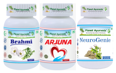
  1. Brahmi Capsules
  2. Arjun Capsules
  3. Neurogenie Capsules

Herbal Remedies of Mother Fixation

Buy Now Herbal Remedies ofMother Fixation

Buy Now Herbal Remedies of Mother Fixation

PRODUCT DESCRIPTION

1.Brahmi Capsules

Formulated with pure, standardized extract of Brahmi (Bacopa monnieri), these capsules serve as an effective brain tonic and nervous system rejuvenator. Brahmi balances the mind, clears mental fog (Tamas), reduces restless Vata energy, and promotes cognitive sharpness, emotional strength, and calm discernment.

Dosage: 1–2 capsules twice daily, with plain water after meals.

2. Arjun Capsules

Prepared from the standardized extract of Arjuna (Terminalia arjuna). While widely known for cardiovascular support, Arjuna in Ayurveda is deeply connected to the physical and subtle heart (Hridaya)—the seat of Ojas and emotional processing. It helps stabilize deep-seated emotional agitation, balances Pitta-Vata disturbances in the heart space, and fosters emotional resilience during stressful transitions.

Dosage: 1–2 capsules twice daily, with plain water after meals.

3. Neurogenie Capsules

A specialized polyherbal formulation combining potent Medhya herbs such as Brahmi (Bacopa monnieri) and Ashwagandha (Withania somnifera). Neurogenie Capsules are engineered to nourish the nervous system (Majja Dhatu), strengthen Manovaha Srotas, reduce anxiety, and improve stress adaptation, supporting mental independence and clarity.

Dosage: 1–2 capsules twice daily, with plain water after meals.

CONCLUSION

Mother fixation is a complex psychological dynamic that hinders an individual’s journey toward emotional autonomy, self-reliance, and fulfilling personal relationships. While its origins lie in unresolved early attachment patterns and family enmeshment, recovering emotional independence is achievable. Modern psychotherapeutic interventions provide structured pathways for establishing boundaries and processing attachment dynamics, while Ayurveda offers a holistic approach by clearing Manovaha Srotas, strengthening Sattva Guna, and balancing internal doshas. Combining psychological therapy, conscious lifestyle adjustments, and supportive Ayurvedic herbal care creates a well-rounded path toward emotional maturity and healthy self-actualization.

Herbal Remedies of Steven Johnson Syndrome – Causes, Symptoms, Diagnosis & Treatment

ABSTRACT

Stevens-Johnson Syndrome (SJS) is a rare but potentially life-threatening mucocutaneous disorder that requires prompt recognition, appropriate evaluation, and comprehensive medical management. It represents a significant clinical challenge because of its acute progression and the possibility of serious complications. Particular attention is given to the importance of early intervention and coordinated supportive care to improve patient outcomes. From an Ayurvedic perspective, SJS can be discussed through the broader concepts of Pitta Dushti, Rakta Dushti, Agni disturbance, and involvement of Twak and Rakta Dhatu.

Steven Johnson Syndrome

STATISTICS FOR STEVEN JOHNSON SYNDROME

  • Approximately 1–5.76 cases per million people per year.
  • May occur at any age; higher rates are reported in children and older adults.
  • Epidermal detachment involves <10% of total body surface area (TBSA).
  • SJS mortality: Approximately 5.4% in isolated SJS.
  • SJS–TEN overlap: Involves 10–30% TBSA, with mortality around 14.4%.
  • TEN: Involves >30% TBSA, with reported mortality of approximately 15.3–50%.

INTERESTING FACTS ABOUT STEVEN JOHNSON SYNDROME

Rare, but memorable

SJS is estimated to affect only a few people per million each year, making it an uncommon disorder.

A medical spectrum

SJS, SJS-TEN overlap, and TEN are regarded as different points along the same disease spectrum.

The 10% rule

The boundary between SJS and SJS-TEN overlap is defined using a surprisingly precise measurement—10% of total body surface area.

A global condition

SJS occurs worldwide, but its frequency and associated risks can vary between populations.

One name, many consequences

Despite being classified as a dermatological disorder, SJS can have implications extending far beyond the skin.

Recovery can be a long journey

The acute episode may resolve, but some individuals require prolonged follow-up and rehabilitation.

lesson in pharmacogenomics

SJS demonstrates how genetic makeup can interact with drug exposure, an important concept in personalized medicine.

The name has a history

The syndrome was first described in 1922 by pediatricians Albert Mason Stevens and Frank Chambliss Johnson.

INTRODUCTION

Stevens-Johnson Syndrome (SJS) is a rare and potentially life-threatening mucocutaneous disorder characterized by extensive epithelial injury and a complex systemic inflammatory response. It is considered part of the same clinical spectrum as toxic epidermal necrolysis (TEN), with the distinction between these entities primarily based on the extent of epidermal detachment. The disorder represents a significant diagnostic and therapeutic challenge because its presentation can evolve rapidly and may involve multiple organ systems. SJS also represents an important area of study in modern medicine because it illustrates the interaction between individual susceptibility, immune-mediated tissue injury, and environmental or therapeutic exposures. Advances in pharmacogenomics have further demonstrated that genetic variation may influence susceptibility in particular populations.

CAUSES AND RISK FACTORS OF STEVEN JOHNSON SYNDROME

The causative relationship is often complex and may involve an interaction between the triggering agent, immune response, and genetic background. These include:

Medication-Related Causes

  • Antibiotics: Particularly sulfonamide antibiotics such as trimethoprim-sulfamethoxazole; other implicated groups include penicillins, cephalosporins, fluoroquinolones, and tetracyclines.
  • Antiepileptic medicines: Carbamazepine, phenytoin, lamotrigine, phenobarbital, and related agents are important recognized triggers.
  • Allopurinol: A well-established high-risk medicine, particularly in genetically susceptible individuals.
  • NSAIDs: Especially the oxicam group, including meloxicam and piroxicam, has been associated with SJS/TEN.
  • Antiretroviral medicines: Nevirapine and certain other antiretroviral agents have been implicated.
  • Other medicines: Immune checkpoint inhibitors and several less frequently implicated drugs have also been reported.

Infectious Triggers

  • Infections can occasionally be associated with SJS, particularly when no responsible medication is identified.
  • Mycoplasma pneumoniae is an important infectious association, particularly in younger patients.
  • Viral infections, including certain herpesviruses and hepatitis viruses, have also been reported.
  • The precise contribution of infection can be difficult to establish because medications are often administered during infectious illnesses.

Genetic Susceptibility

  • HLA (human leukocyte antigen) variation is an important determinant of susceptibility to drug-associated SJS/TEN.
  • HLA-B*15:02 is strongly associated with carbamazepine-related SJS/TEN in several Asian populations, including specific Indian populations.
  • HLA-B*58:01 is associated with increased susceptibility to allopurinol-induced SJS/TEN.
  • HLA-A*31:01 has been associated with carbamazepine-related reactions in several populations.
  • These associations are drug- and population-specific, rather than indicating a universal genetic risk.

Immunological and Host Factors

  • SJS/TEN represents a T-cell-mediated delayed hypersensitivity reaction.
  • An altered immune state may increase susceptibility to severe cutaneous adverse reactions.
  • Individuals with HIV infection have a substantially increased risk compared with the general population.
  • Malignancy, autoimmune disorders, and other states of immune dysfunction have also been reported as associated factors.

Other Recognized Risk Factors

  • Previous exposure to a culprit medicine may be relevant, particularly when re-exposure occurs.
  • Multiple concomitant medicines can make identification of the responsible agent difficult.
  • Kidney or liver dysfunction may be associated with increased vulnerability in some patients and can influence drug handling.
  • Certain age groups, particularly young children and older adults, have been reported to have increased risk in epidemiological studies.

CLINICAL FEATURES OF STEVEN JOHNSON SYNDROME

It is characterized by an acute, rapidly evolving mucocutaneous illness. Its clinical picture may vary between patients, but several characteristic findings help distinguish it from other severe cutaneous disorders. These include:

Acute Onset

The condition generally develops abruptly, with rapid progression over a short period. A preceding period of systemic illness may occur before prominent mucocutaneous manifestations become evident.

Prodromal Illness

Patients may initially develop fever, malaise, fatigue, and generalized discomfort. Headache, arthralgia, or other nonspecific constitutional complaints may accompany the early phase.

Skin Lesions

Cutaneous involvement commonly begins with erythematous or dusky macules. Lesions may develop an atypical targetoid or concentric appearance. The lesions can become confluent as the disease progresses. Atypical target lesions are particularly characteristic of SJS.

Epidermal Detachment

Progressive injury to the epidermis can result in flaccid blister formation and epidermal separation. Gentle mechanical pressure may cause superficial epidermal sloughing, a phenomenon known as positive Nikolsky sign. In SJS, epidermal detachment typically involves less than 10% of total body surface area.

Ocular Features

Findings can include conjunctivitis, ocular surface epithelial defects, and corneal involvement. Severe ocular disease may subsequently result in scarring, dry eye, visual impairment, or other chronic complications.

Systemic Involvement

SJS is not restricted to the skin and mucous membranes. Systemic involvement may include the respiratory, gastrointestinal, renal, hepatic, and hematological systems.

Genitourinary Manifestations

Urogenital mucosal involvement may lead to painful urination and difficulty passing urine.

DIAGNOSIS OF STEVEN JOHNSON SYNDROME

The diagnosis is primarily based on clinical assessment, supported by a detailed history, physical examination, and appropriate investigations. Since no single laboratory test can definitively establish SJS, careful evaluation of the temporal relationship with potential triggers, characteristic mucocutaneous findings, and the extent of epidermal involvement is essential.

Detailed medication history

SJS is primarily diagnosed from the clinical history and characteristic mucocutaneous findings. Review all recently started, stopped, or dose-adjusted medicines, particularly those taken during the preceding several weeks.

Physical examination

Assess the skin, oral cavity, eyes, genital region, and other mucosal surfaces systematically. 

Extent of epidermal detachment

Estimate the percentage of total body surface area (TBSA) showing epidermal detachment. SJS generally involves <10% TBSA.

Skin biopsy

A punch or incisional biopsy from an appropriate lesion can support the diagnosis and help distinguish SJS/TEN from other blistering disorders. Typical findings include epidermal necrosis with separation of the epidermis from the underlying dermis and relatively limited inflammation.

Laboratory evaluation

Complete blood count, serum electrolytes, renal and liver function tests, glucose, and other investigations help assess systemic involvement and disease severity.

Multidisciplinary evaluation

Severe cases benefit from coordinated assessment involving dermatology, ophthalmology, critical care, internal medicine, and other relevant specialties.

MANAGEMENT OF STEVEN JOHNSON SYNDROME

It requires prompt recognition, immediate clinical assessment, and coordinated supportive care because of its potentially rapid and severe course. The management approaches include:

Immediate Withdrawal of the Suspected Trigger

Identify and promptly discontinue the suspected causative medicine. Non-essential medicines should also be reviewed and stopped when clinically appropriate.

Fluid and Electrolyte Management

Extensive epidermal injury can cause substantial fluid and electrolyte losses. Monitor blood pressure, urine output, electrolytes, renal function, and fluid balance regularly.

Skin and Wound Care

Use non-adherent or petrolatum-impregnated dressings to protect denuded areas. Detached epidermis may be preserved as a biological covering when appropriate.

Pain Management

Appropriate analgesia should be provided according to pain severity. Severe pain may require opioid analgesics under monitored hospital care.

Nutritional Support

Adequate nutritional intake is essential for epithelial repair and recovery. If oral intake is inadequate because of mucosal involvement, enteral nutritional support may be considered.

Assessment of Disease Severity

SCORTEN can be used in appropriate patients to estimate mortality risk. Relevant parameters include age, malignancy, heart rate, extent of epidermal detachment, serum urea, glucose, and bicarbonate.

Immunomodulatory Therapy

Immunomodulators like cyclosporine, systemic corticosteroids, intravenous immunoglobulin, TNF-alpha inhibitors such as etanercept can be given.

AYURVEDA INSIGHTS

Its manifestations may be conceptually understood through the interplay of Pitta Dosha, Rakta Dhatu, Twak, Agni, and Srotas, particularly where inflammatory processes and tissue injury are prominent. Pitta aggravation and Rakta Dushti may be considered relevant to the pathological expression, while impaired Agni and systemic Dosha-Dhatu disturbance may contribute to disease progression. Ayurvedic assessment would therefore emphasize individual constitution, Dosha status, Agni, and Dhatu involvement.

PATHOGENESIS OF STEVEN JOHNSON SYNDROME

From an Ayurvedic conceptual standpoint, the pathogenesis of Stevens-Johnson Syndrome may be explored as a complex disturbance of Dosha, Dushya, and Srotas, rather than being assigned to a single classical disease. The process may begin with aggravation of Pitta, particularly its association with Rakta, followed by impairment of normal tissue metabolism and disturbance of the body’s internal balance. When aggravated Doshas interact with Rakta and Twak, the integrity and nourishment of these tissues may become progressively compromised. Disturbance of Rakta Vaha Srotas may further facilitate the spread of the pathological process, while weakened Agni can impair proper transformation and tissue nourishment. In an Ayurvedic conceptual interpretation of SJS, the Dosha–Dushya sequence can be presented as follows:

  • Dosha: Predominant Pitta aggravation, particularly involving its association with Rakta, initiates the pathological imbalance.
  • Dushya: The aggravated Dosha interacts primarily with Rakta and Twak, which may be regarded as the principal affected tissues in this conceptual model.
  • Srotas: Continued Dosha-Dushya interaction may disturb Rakta Vaha Srotas, facilitating further tissue involvement.
  • Adhisthana: Twak and associated superficial tissues may be considered the principal sites where the pathological process becomes established.
  • Vyakti: The accumulated pathological changes eventually become clinically apparent as the characteristic manifestations of the condition.

HERBAL REMEDIES FOR STEVEN JOHNSON SYNDROME BY PLANET AYURVEDA

Planet Ayurveda is a well-established Ayurvedic healthcare organization that works to promote the ancient knowledge of Ayurveda through natural remedies and holistic health solutions. The company develops a wide range of herbal formulations using pure, high-quality ingredients and standardized herbal extracts, with a focus on maintaining safety, authenticity, and effectiveness. Following the principles of classical Ayurveda, Planet Ayurveda aims to support overall health by addressing the underlying causes of health concerns and encouraging balance in the body, mind, and emotions. With an emphasis on quality, natural ingredients, and traditional approaches supported by modern research, the organization works toward making Ayurvedic wellness solutions available globally. We will explore various herbal approaches used for the management of Steven Johnson Syndrome  according to Ayurvedic principles. The herbal formulations are:

  1. Kaishore Guggul
  2. Gandhak Rasayan
  3. Manjishtha Capsules
  4. Gleaming Skin Hair Nails Formula
  5. Chopchini Churna

PRODUCT DESCRIPTION

1. Kaishore Guggul

It is a classical Ayurvedic formulation containing Amalaki (Emblica officinalis), Bibhitaki (Terminalia bellirica), Haritaki (Terminalia chebula), Guggulu (Commiphora mukul), Guduchi (Tinospora cordifolia), Ginger (Zingiber officinale), Black Pepper (Piper nigrum), Long Pepper (Piper longum), Vidanga (Embelia ribes), Red Physic Nut (Baliospermum montanum), and Indian Jalap (Ipomoea turpethum). Traditionally, it is used in Pitta-Rakta disorders, inflammatory conditions, skin disorders, and metabolic disturbances associated with Ama. Its classical properties may support Rakta Shodhana, reduction of inflammatory burden, and restoration of Dosha balance.

Dosage: 2 tablets twice a day with warm water.

2. Gandhak Rasayan

It is prepared with Shuddha Gandhak (purified sulfur), is traditionally regarded in Ayurveda as a formulation supporting Twak, Rakta, and Pitta balance and maintaining healthy tissue function. In the context of Stevens-Johnson Syndrome, its possible role may be considered only from an Ayurvedic supportive perspective, particularly during the recovery phase. Traditionally, purified sulfur is associated with Kushthaghna, Krimighna, and Rasayana properties and may support skin tissue nourishment and recovery.

Dosage: 2 tablets twice a day with plain water.

3. Manjishtha Capsules

These contain Manjishtha (Rubia cordifolia), an Ayurvedic herb traditionally valued for its influence on Rakta Dhatu and skin health. In an Ayurvedic supportive framework for Stevens-Johnson Syndrome, Manjishtha may be considered for its traditional Rakta Shodhaka, Pitta-pacifying, and Twak-supportive properties. Its phytoconstituents have been investigated for antioxidant and anti-inflammatory activities, which may provide a rationale for supporting tissue recovery after the acute inflammatory phase. It may therefore be considered during post-acute recovery to support restoration of skin and tissue health.

Dosage: 1 capsule twice a day after meals with plain water.

4. Gleaming Skin Hair Nails Formula

These contain Manjishtha (Rubia cordifolia), Pit Papda (Fumaria officinalis), Ghritkumari (Aloe barbadensis), and Sariva (Hemidesmus indicus). Traditionally, these herbs are valued for supporting Rakta Dhatu, Twak health, and tissue nourishment. In an Ayurvedic supportive approach to Stevens-Johnson Syndrome, the formulation may be considered during the post-acute recovery phase, when the focus shifts toward restoration of skin integrity and overall tissue health. Manjishtha and Sariva are traditionally regarded as Rakta-prasadana herbs, while Ghritkumari is valued for its soothing and tissue-supportive properties.

Dosage: 1 capsule twice a day after meals with plain water.

5. Chopchini Churna

It contains the rhizome powder of Chopchini (Smilax china), traditionally used in Ayurveda for supporting Rakta Dhatu and skin health. Its classical properties are considered useful in conditions associated with inflammatory imbalance and impaired tissue health. In an Ayurvedic supportive approach to Stevens-Johnson Syndrome, Chopchini may be considered during the recovery phase to support tissue nourishment and restoration of healthy skin function. Its traditional therapeutic profile may complement efforts to maintain Dosha balance and Rakta Dhatu integrity.

Dosage: ½ teaspoon twice a day.

CONCLUSION

Stevens-Johnson Syndrome requires a carefully coordinated approach that extends beyond management of the acute clinical episode. Successful care depends on close observation, individualized decision-making, and attention to the patient’s physical and functional recovery. Recovery should therefore be viewed as a gradual process requiring continuity between acute care, specialist follow-up, and rehabilitation. From an integrative perspective, Ayurvedic principles may provide a conceptual framework for individualized supportive care during suitable recovery stages, particularly with attention to Agni, Dosha balance, Rakta, and Dhatu nourishment.

Herbal Remedies of Cocoon Syndrome – Causes, Symptoms, Diagnosis & Treatment

ABSTRACT

Cocoon Syndrome, also known clinically as abdominal cocoon, is a rare and unusual condition characterized by the partial or complete encasement of the small intestine within a dense, fibrous, collagenous membrane resembling a cocoon. This mechanical entrapment frequently leads to acute or chronic intestinal obstruction, recurrent abdominal pain, nausea, vomiting, and malnutrition. While conventional management often relies on exploratory laparotomy and surgical membranectomy, patients frequently seek complementary and holistic approaches to reduce systemic inflammation, prevent tissue fibrosis, and enhance gastrointestinal motility.

In Ayurveda, structural entrapments and motility disorders of the abdomen are interpreted through the lens of Annavaha Srotas (gastrointestinal system) dysfunction, involving the severe aggravation of Vata and Kapha doshas, deep-seated metabolic impurities (Ama), and localized tissue hardening or obstruction (Sanga). Ayurvedic management focuses on Vata-Kapha pacifying therapies (Samshodhana and Samshamana), deep tissue detoxification, and formulations that promote carminative action (Deepana-Pachana), relieve bowel congestion, and restore normal downward movement of energy (Anulomana). This article explores the comprehensive causes, clinical manifestations, diagnostic approaches, conventional interventions, and the targeted Ayurvedic management of cocoon syndrome using specific herbal formulations

 Cocoon Syndrome

INTRODUCTION

Cocoon Syndrome represents a challenging clinical entity within gastroenterology and surgery, characterized by a fibro-collagenous sac enveloping loops of the small intestine. This anatomical encasement restricts normal peristalsis and can cause recurrent episodes of subacute or complete intestinal obstruction. Patients often present with colicky abdominal pain, recurrent vomiting, weight loss, and a palpable abdominal mass.

The pathogenesis of cocoon syndrome is often categorized as primary (idiopathic, frequently observed in adolescent females residing in tropical or subtropical regions) or secondary (linked to continuous ambulatory peritoneal dialysis, beta-blocker therapy, peritoneal ventriculoperitoneal shunts, or abdominal tuberculosis). While surgical adhesiolysis and membranectomy remain the cornerstone of conventional treatment, high rates of postoperative recurrence or persistent dysmotility necessitate supportive and corrective medical care.

Integrating Ayurvedic principles into the care continuum offers a holistic perspective aimed at clearing metabolic toxins, melting deep-seated fibrous accumulations, regulating digestive fire (Agni), and restoring normal intestinal transit.

CAUSES

The etiology of cocoon syndrome spans idiopathic, environmental, iatrogenic, and infectious factors:

  • Primary (Idiopathic) Factors: Often seen in young adolescent girls in tropical climates, potentially linked to retrograde menstruation or subclinical viral peritonitis.
  • Secondary Causes: Associated with long-term peritoneal dialysis, abdominal tuberculosis, sarcoidosis, or gynecological conditions.
  • Pharmaceutical Agents: Prolonged usage of specific medications like practolol has historically been documented to induce sclerosing peritonitis.
  • Chronic Abdominal Inflammation: Persistent low-grade inflammation of the peritoneum leading to excessive collagen deposition and membrane formation.

SIGNS  AND SYMPTOMS

The clinical presentation of cocoon syndrome is often intermittent or acute depending on the degree of bowel strangulation:

  • Recurrent Colicky Abdominal Pain: Cramping pain resulting from obstructed peristalsis within the fibrous sac.
  • Abdominal Distension and Nausea: Noticeable bloating and frequent postprandial nausea due to delayed gastric emptying and transit.
  • Palpable Abdominal Mass: A soft, non-tender tumor-like mass felt during physical examination, representing the encapsulated bowel loops.
  • Malnutrition and Weight Loss: Chronic malabsorption and fear of eating due to post-meal pain leading to significant nutritional deficiency.
  • Vomiting: Bilious or non-bilious vomiting occurring during acute exacerbations of intestinal obstruction.

INVESTIGATIONS

Accurate anatomical visualization and exclusion of other causes of bowel obstruction are vital:

  • Plain Abdominal Radiography: May reveal clustered small bowel loops, dilated air-fluid levels, or calcifications within the sac.
  • Contrast-Enhanced Computed Tomography (CECT): Considered the gold standard imaging modality, demonstrating a “cauliflower-like” clustering of small bowel loops enclosed within a thick, enhancing peritoneal membrane.
  • Barium Studies: Upper gastrointestinal series with small bowel follow-through showing delayed transit and fixed, huddled bowel loops.
  • Diagnostic Laparoscopy or Laparotomy: Direct visualization of the dense fibro-collagenous membrane encasing the intestine, confirming the final diagnosis.

MANAGEMENT

Conventional management of cocoon syndrome primarily involves surgical intervention for symptomatic or obstructed cases.

  • Conservative Measures: Bowel rest, nasogastric decompression, total parenteral nutrition (TPN), and close monitoring for acute ischemic changes in mild or early presentations.
  • Surgical Intervention: Open exploratory laparotomy with careful peeling and excision of the thick encapsulating membrane (membranectomy) along with extensive lysis of adhesions to free the entrapped small bowel loops.
  • Supportive Medical Care: Postoperative management focusing on gut rehabilitation, anti-inflammatory support, and herbal protocols to restore regular bowel habits and prevent recurrence.

AYURVEDIC FORMULATIONS FOR GASTROINTESTINAL HEALTH

Planet Ayurveda provides authentic, high-quality herbal formulations crafted in GMP-certified facilities using standardized extracts. Free from synthetic additives, chemicals, and heavy metals, these products support holistic digestive recovery and metabolic balance.

The specific protocol suggested for the management of Cocoon Syndrome includes:

  1. Triphala capsules
  2. Panchsakar churna
  3. Syrup Digestoplan
  4. Syrup Acidoplan
  5. Mahashankh vati

Herbal Remedies of Cocoon Syndrome

Buy Now Herbal Remedies of Cocoon Syndrome

Buy Now Herbal Remedies of Cocoon Syndrome

PRODUCT DESCRIPTION

1.  Triphala Capsules

Formulated with a balanced blend of three classical fruits: Terminalia chebula (Haritaki), Terminalia bellirica (Bibhitaki), and Emblica officinalis (Amalaki). Triphala is a legendary Ayurvedic rasayana renowned for gentle internal cleansing, detoxification, and regulation of bowel movements. It helps clear accumulated toxins (Ama) from the gastrointestinal tract, supports healthy mucosal lining, and promotes regular peristalsis without causing dependency.

Dosage: 1 capsule twice daily, with plain water after meals.

2. Panchsakar Churna

A traditional herbal powder combining five potent ingredients: Senna, Salt of Haritaki, Shunti (Ginger), Sauvatchal Lavana (Black Salt), and Trivrit. Panchsakar Churna is exceptionally effective in clearing bowel congestion, relieving abdominal heaviness, and maintaining proper digestive elimination. It acts as a natural carminative that alleviates gas, bloating, and sluggish bowel transit.

Dosage: 1/2 teaspoon twice daily, with warm water.

3. Syrup Digestoplan        

A comprehensive herbal syrup designed to strengthen digestion and improve nutrient assimilation. It stimulates the secretion of digestive enzymes, balances gastric acid production, and relieves post-meal discomfort, nausea, and abdominal distension associated with compromised gut motility.

Dosage: 2 teaspoons twice daily, after meals.

4. Syrup Acidoplan

Formulated to soothe the gastrointestinal tract and balance Pitta and Vata doshas in the upper digestive tract. Syrup Acidoplan helps reduce burning sensations, acidity, and irritation in the stomach lining, providing a calming environment for sensitive digestive tissues undergoing rehabilitation.

Dosage: 2 teaspoons twice daily, after meals.

5. Mahashankh Vati

A classic mineral-herbal Ayurvedic tablet containing potent digestive and carminative substances like Shankh Bhashma, Danti, Pippali, and various salts. Mahashankh Vati is highly regarded for managing chronic indigestion, abdominal colic, gas accumulation, and sluggish metabolism. It enkindles digestive fire (Agni) and helps dissolve deep-seated metabolic obstructions.

Dosage: 2 tablets twice daily to be chewed 20 minutes before meals.

CONCLUSION

Cocoon Syndrome is a rare and complex gastrointestinal condition characterized by small bowel encasement and obstruction, significantly impacting a patient’s nutritional status and quality of life. While conventional surgery is often required for mechanical release, integrating traditional Ayurvedic principles offers a valuable supportive approach for long-term gut rehabilitation. By addressing underlying doshic imbalances, clearing metabolic toxins (Ama), reducing fibrous tissue accumulation, and enhancing digestive fire through targeted herbal protocols—such as Triphala capsules, Panchsakar churna, Syrup Digestoplan, Syrup Acidoplan, and Mahashankh vati—patients can achieve better digestive health, improved motility, and enhanced overall well-being.

Herbal Remedies of Depleted Dad Syndrome – Causes, Symptoms, Diagnosis & Treatment

ABSTRACT

Modern life demands a continuous balancing act between professional responsibilities, family caregiving, and everyday obligations, often leaving individuals with a sense of persistent exhaustion that a simple night of sleep cannot resolve. Among fathers, this manifests as “Depleted Dad Syndrome”—a descriptive term capturing the broader experience of parental burnout when ongoing demands repeatedly outpace a person’s recovery resources. While not an established medical diagnosis, this state of chronic physical, mental, and emotional depletion affects energy, patience, concentration, and enthusiasm. While modern psychology and occupational frameworks help categorize burnout, Ayurveda views this chronic fatigue through the lens of Vata aggravation, Agni imbalance, and the depletion of vital energy (Ojas) resulting from excessive mental preoccupation (Ati-Chinta). In this article, we shall explore the causes, warning signs, clinical considerations, and comprehensive management of Depleted Dad Syndrome through both modern and Ayurvedic perspectives.

Depleted Dad Syndrome

INTRODUCTION

Depleted Dad Syndrome describes a state of persistent physical, mental, and emotional exhaustion experienced by fathers when ongoing responsibilities repeatedly exceed available recovery resources. Unlike ordinary tiredness after a busy workday, the core difficulty lies in the feeling of never completely recharging.

Modern fatherhood often involves juggling multiple overlapping roles simultaneously-acting as a professional, provider, partner, parent, caregiver, decision-maker, and problem-solver. Consequently, the mind remains in a continuous “responsibility mode,” where work extends past office hours into financial planning, household decisions, and future concerns. When adequate sleep, nutrition, physical activity, and emotional recovery are missing, the accumulated load becomes difficult to manage, highlighting that parental burnout is not solely a maternal issue but a significant challenge for fathers as well

CAUSES AND CONTRIBUTING FACTORE

Depleted Dad Syndrome arises when the continuous demands of modern life and family responsibilities chronically outbalance personal recovery resources.

Common Contributors Include

  • Overlapping Roles and Responsibilities: Juggling professional duties, financial planning, parenting, and household decisions without adequate mental breaks.
  • Continuous Responsibility Mode: The inability to switch off mentally, keeping the brain preoccupied with deadlines, family needs, and future planning even during rest.
  • Chronically Unbalanced Demand and Recovery: Operating under high pressure while sacrificing sleep, proper nutrition, physical activity, and recreation.
  • Parenting Stress: Navigating the complex emotional and practical demands of raising children, which research links closely to parental burnout and reduced parental efficacy.

RISK FACTORS INCLUDE

  • High-pressure career environments with demanding work hours or blurred boundaries between work and home.
  • Limited external social support networks or lack of personal time for hobbies and recreation.
  • Perfectionistic tendencies or an internalized pressure to constantly provide and solve problems independently.
  • Poor sleep hygiene or chronic sleep deprivation due to family or work obligations.
  • Lack of structured transitions between professional duties and family life.

SIGNS AND SYMPTOMS

The manifestations of Depleted Dad Syndrome build up gradually, affecting personal well-being, mental focus, and family interactions.

Common Symptoms Include

  • Persistent Tiredness or Low Energy: Waking up tired and experiencing the frustrating feeling of sleeping yet still feeling exhausted.
  • Mental Overload and Reduced Concentration: Feeling mentally foggy, finding it difficult to sustain attention on a single task, or experiencing persistent thoughts about work and responsibilities.
  • Irritability and Shorter Temper: Noticeable drops in patience where small inconveniences feel disproportionately demanding.
  • Emotional Withdrawal: Seeking isolation, silence, or screen time due to having little emotional energy left for active family participation.
  • Loss of Enthusiasm: Gradual disappearance of hobbies, exercise, social interactions, or other activities that once provided relaxation.
  • Feeling Permanently “On Duty”: The inability to feel completely off-duty, with the mind constantly preparing for the next responsibility even during rest.
  • Reduced Sense of Accomplishment: Feeling that responsibilities are continuously piling up despite working extremely hard.

INVESTIGATIONS AND CLINICAL EVALUATION

Persistent exhaustion deserves careful attention to rule out underlying medical conditions and distinguish burnout from other health concerns.

Clinical Assessment

  • Detailed Health and Lifestyle History: Examining sleep patterns, work demands, family responsibilities, and daily routines to identify chronic imbalances.
  • Exclusion of Medical Conditions: Checking for physiological causes of fatigue, such as sleep disorders, anemia, thyroid disorders, nutritional deficiencies, metabolic disease, medication side effects, and mood disorders.
  • Psychological Evaluation: Assessing for overlapping conditions emphasized in clinical literature, such as depression, anxiety, and other primary causes of exhaustion.

Differential Screening & Diagnostics

  • Sleep Studies: Evaluating for underlying sleep disorders if chronic fatigue persists despite adequate time in bed.
  • Blood and Metabolic Panels: Screening for biomarkers related to anemia, thyroid function, and nutritional status.
  • Burnout vs. Mood Disorder Assessment: Distinguishing the dimensions of burnout (energy depletion, mental distance, reduced efficacy) from clinical depression or generalized anxiety.

MANAGEMENT

Managing Depleted Dad Syndrome focuses on restoring balance between demands and recovery resources rather than adding more tasks to an already overloaded schedule.

Conventional and Lifestyle Approaches Include

  • Establishing Consistent Sleep Schedules: Treating sleep as a non-negotiable biological requirement rather than a flexible resource to sacrifice for work.
  • Regular Nutrition: Avoiding skipped meals and heavy reliance on caffeine or highly processed foods to maintain stable daily energy.
  • Daily Movement: Incorporating walking, yoga, strength training, or other appropriate exercise forms into the routine to aid recovery.
  • Creating Work-Home Transitions: Using short walks, quiet tea, breathing practices, or 10–15 minutes without digital stimulation to build a psychological boundary between roles.
  • Protecting Personal Time: Recognizing that rest and personal space for reflection and recreation are essential components of sustainable fatherhood.

AYURVEDIC VIEW AND MANAGEMENT

Ayurveda views prolonged stress and inadequate recovery through traditional frameworks involving Ati-Chinta (excessive mental preoccupation), Vata aggravation, Agni imbalance, and the depletion of Ojas (vitality and resilience). A high-demand lifestyle disrupts Vata and disturbs sleep and mental rest, which in turn impacts digestion, tissue nourishment, and overall strength (Daurbalya).

Ayurvedic management focuses on restoring the foundation through proper diet (Ahara), sleep (Nidra), lifestyle routines (Vihara), mental balance (Manasika balance), and appropriate herbal support, rather than relying on stimulant-based quick fixes.

HOME REMEDY: SUPPORTIVE DAILY ROUTINE & NUTRITION

Establishing grounding daily habits helps calm Vata and support mental clarity without adding complexity to the day.

Principles of the Approach

  • Regular Meal Times: Eating fresh, warm, nourishing meals at consistent times to support Agni (metabolic fire).
  • Wind-Down Practices: Setting aside digital devices before bed to encourage deeper, more restorative sleep (Nidra).
  • Mindful Transitions: Taking brief pauses throughout the day to reset mental focus and reduce accumulation of stress.
  • Nasya (calming)– 2-2 drops of lukewarm desi ghee in each nostril.
  • Paadabhyang (foot massage)- Daily foot massage with warm Sesame oil  will help in improving the mind’s exhaustion.

Herbal Remedies for Depleted Dad Syndrome by Planet Ayurveda

Planet Ayurveda is a globally recognized healthcare organization dedicated to offering authentic Ayurvedic formulations manufactured in GMP-certified facilities. Prepared using standardized herbal extracts, Planet Ayurveda’s products are free from synthetic additives, chemicals, and heavy metals. By blending traditional Ayurvedic wisdom with modern quality controls, Planet Ayurveda provides herbal supplements designed to support daily wellness, vitality, and balance.

Planet Ayurveda presents the following list of products to support cardiovascular wellness, male vitality, and nervous system health:

  1. Arjun Tea
  2. Tribulus Power Capsules
  3. NeuroGin Capsules

Herbal Remedies of Depleted Dad Syndrome

Buy Now Herbal Remedies of Depleted Dad Syndrome

Buy Now Herbal Remedies of Depleted Dad Syndrome

PRODUCT DESCRIPTION

1. Arjun Tea

A herbal tea formulated to support healthy circulation, cardiovascular function, and daily metabolic wellness. It contains Arjuna (Terminalia arjuna), Punarnava (Boerhavia diffusa), Pipal Tvak (Ficus religiosa), Dalchini (Cinnamomum zeylanicum), and Chhoti Elaichi (Elettaria cardamomum). In Ayurveda, Arjuna is regarded as a Hridya herb, making this tea a heart-supportive daily beverage rather than a stimulant-based energy drink.

2. Tribulus Power Capsules

A vegetarian capsule preparation based on standardized extract of Gokshura (Tribulus terrestris). In Ayurveda, Gokshura is described with properties including Vrishya (vitality-promoting), Brihana (nourishing), and Tridosha-supportive actions, contributing to a broader approach to physical and male vitality.

3. NeuroGin Capsules

A specialized formulation combining standardized extracts of Brahmi (Bacopa monnieri) and Ashwagandha (Withania somnifera) at 250 mg each per capsule. Brahmi is associated with Medhya functions (supporting intellect and mental clarity), while Ashwagandha is regarded as a Rasayana (rejuvenating)  and Balya herb associated with strength, nourishment, and resilience, providing support for the mind and nervous system during periods of sustained mental workload.

CONCLUSION

Depleted Dad Syndrome captures the modern reality of fathers who continue fulfilling heavy responsibilities while their physical, mental, and emotional reserves gradually run low. While not an official medical diagnosis, parental burnout and chronic stress deserve thoughtful attention and care. Ayurveda provides a comprehensive lens through Vata balance, Agni (digestive fire), nourishment, Nidra (sleep), and Ojas (vitality and resilience) to understand and address exhaustion. Under professional guidance, incorporating lifestyle adjustments and supportive formulations like Planet Ayurveda Arjun Tea, Tribulus Power, and NeuroGin can help a father recover his reserves, restore his energy, and remain physically present, mentally clear, and emotionally available for his family.


Herbal Remedies of Urinary Tract Infection – Causes, Symptoms, Diagnosis & Treatment

ABSTRACT

Urinary Tract Infection (UTI) is a condition that deserves attention because even a seemingly minor urinary complaint can sometimes develop into a more serious problem if ignored. Changes in urination, burning sensation, pelvic discomfort, urgency, or unusual urine appearance may indicate an underlying urinary infection. Women commonly experience UTIs, but no age group is completely protected. Maintaining adequate fluid intake, healthy personal hygiene, and proper urinary habits can help reduce the risk. Today, I would like to tell you about UTI in detail, including why it occurs, its common signs and symptoms, contributing factors, diagnostic methods, possible complications, and how it can be understood and managed through both modern medicine and Ayurveda.

 Urinary Tract Infection

INTRODUCTION

Urinary Tract Infection (UTI) is one of the most common infections affecting the urinary system and can involve the urethra, urinary bladder, ureters, or kidneys. It occurs when microorganisms, most commonly bacteria, enter and multiply within the urinary tract, leading to symptoms such as burning during urination, frequent or urgent urination, lower abdominal discomfort, cloudy or foul-smelling urine, and sometimes fever. Although UTIs can affect people of all age groups, they are particularly common among women. Recurrent or inadequately managed infections may progress to more serious conditions, including kidney involvement and systemic infection.

STATISTICS OF URINARY TRACT INFECTION

Urinary Tract Infection (UTI) is a widespread health problem, particularly among women. . More than 50% of adult women experience at least one UTI during their lifetime. Nearly 25% of women who develop a bladder infection may experience another episode, making recurrence a common concern. Women are affected much more frequently than men, largely due to anatomical differences that allow bacteria to reach the urinary tract more easily. UTI is one of the most commonly diagnosed bacterial infections in community and healthcare settings and contributes significantly to antibiotic use. The risk of UTI may increase with advancing age, especially when factors such as incomplete bladder emptying, urinary obstruction, or catheter use are present. Together, these statistics demonstrate the substantial burden of UTI and emphasize the importance of timely diagnosis, appropriate treatment, and preventive urinary care.

INTERESTING FACT ABOUT URINARY TRACT INFECTION

  • UTI can affect anyone: Although more common in women, urinary infections can occur in men, children, and older adults.
  • Bacteria are the usual cause: Most UTIs develop when bacteria from the surrounding area enter the urinary tract and multiply.
  • The bladder is commonly involved: Lower urinary tract infections often affect the bladder and are commonly referred to as cystitis.
  • Some UTIs have few symptoms: Certain people, particularly older adults, may not experience the typical burning or urinary urgency associated with infection.
  • Sexual activity can increase risk: In some women, sexual activity may facilitate the movement of bacteria toward the urethra.
  • Catheters can raise the risk: Urinary catheters provide a pathway through which microorganisms can enter the urinary system.
  • Kidney infections can develop: An untreated or complicated infection may travel from the bladder to the kidneys and require prompt medical attention.
  • Diagnosis is not based only on symptoms: Urine testing, particularly urinalysis and urine culture when indicated, can help identify infection and guide appropriate treatment.

CAUSES OF URINARY TRACT INFECTION

  • Bacterial entry: Most UTIs occur when bacteria from the skin or bowel region enter the urethra and multiply within the urinary tract.
  • Poor personal hygiene: Inadequate hygiene around the genital and urinary area can facilitate the transfer of microorganisms toward the urethra.
  • Insufficient fluid intake: Drinking too little water may result in less frequent urination, reducing the natural flushing of microorganisms from the urinary tract.
  • Holding urine for long periods: Repeatedly postponing urination can interfere with normal bladder emptying and may contribute to bacterial growth.
  • Sexual activity: Sexual intercourse can move bacteria toward the urethral opening, increasing the likelihood of infection in susceptible individuals.
  • Incomplete bladder emptying: Conditions that prevent the bladder from emptying completely can leave residual urine, creating an environment favorable for microbial multiplication.
  • Urinary obstruction: Kidney stones, enlarged prostate, or other blockages can interfere with urine flow and increase infection risk.
  • Urinary catheter use: Catheters can introduce microorganisms into the urinary tract and are an important risk factor for healthcare-associated UTIs.
  • Pregnancy: Physiological and hormonal changes during pregnancy can affect urinary flow and increase susceptibility to UTI.
  • Underlying health conditions: Diabetes and certain disorders affecting the urinary system or immune function can make a person more vulnerable to infection.

SIGN AND SYMPTOMS OF URINARY TRACT INFECTION

  • Burning while urinating: A burning, stinging, or painful sensation may occur during passage of urine.
  • Frequent urination: A person may feel the need to urinate more often than usual, even when only a small amount of urine is passed.
  • Sudden urinary urgency: There may be a strong and difficult-to-control urge to urinate.
  • Lower abdominal discomfort: Pressure, heaviness, cramps, or pain may be felt around the lower abdomen or pelvic region.
  • Changes in urine: Urine may appear cloudy or occasionally contain blood. An unusual or strong odor can also occur.
  • Incomplete emptying: Some individuals may feel that the bladder has not emptied completely after urination.
  • Fever and chills: These symptoms may occur when the infection is more significant or has involved the upper urinary tract.
  • Back or side pain: Pain in the lower back or side, particularly along with fever, may indicate possible kidney involvement.
  • Nausea or vomiting: These symptoms can accompany an upper urinary tract infection, especially when the kidneys are affected.
  • General weakness: Fatigue, reduced appetite, or a feeling of being unwell may accompany a more severe infection

DIAGNOSIS OF URINARY TRACT INFECTION

  • Medical history: The healthcare professional asks about urinary symptoms, their duration, previous infections, medications, pregnancy status, and relevant medical conditions.
  • Physical examination: An examination may be performed to identify abdominal, pelvic, flank, or other findings that could suggest urinary infection or another condition.
  • Urinalysis: A urine sample is examined for findings such as white blood cells, red blood cells, nitrites, and other indicators that may support the presence of infection.
  • Urine culture: The sample is grown in a laboratory to identify the causative microorganism and determine which antibiotics are likely to be effective. This is particularly useful in recurrent, complicated, or suspected upper urinary tract infections.
  • Urine microscopy: Microscopic examination can detect pus cells, bacteria, blood, and other urinary abnormalities that may help in evaluation.
  • Blood investigations: When a severe or upper urinary tract infection is suspected, tests such as a complete blood count and kidney function tests may be recommended.
  • Imaging studies: Ultrasound, CT, or other imaging may be considered when kidney stones, urinary obstruction, structural abnormalities, recurrent infections, or complications are suspected.
  • Pregnancy testing: In women of reproductive age, pregnancy assessment may be appropriate because pregnancy changes the evaluation and management of UTI.
  • Differential diagnosis: Similar symptoms can occur with vaginal infections, sexually transmitted infections, kidney stones, or bladder conditions, so these possibilities may need to be excluded.
  • Clinical assessment: The final diagnosis is made by combining the patient’s symptoms, examination findings, and appropriate laboratory investigations rather than relying on a single test alone.

MANAGEMENT OF URINARY TRACT INFECTION

  • Medical evaluation: Treatment should begin with proper assessment to determine whether the infection is uncomplicated, recurrent, or associated with kidney involvement or another underlying problem.
  • Appropriate antibiotics: When a bacterial UTI is diagnosed, antibiotics may be prescribed according to the type of infection, patient factors, local resistance patterns, and, when required, urine culture results.
  • Adequate hydration: Maintaining sufficient fluid intake can support normal urine production and help prevent dehydration. Fluid intake should be individualized in people with kidney, heart, or other conditions requiring fluid restriction.
  • Regular bladder emptying: Avoiding unnecessary or prolonged postponement of urination may support normal bladder function.
  • Pain and fever management: Suitable medicines may be recommended by a healthcare professional to relieve discomfort, fever, or urinary symptoms.
  • Identify underlying causes: Recurrent infections may require evaluation for factors such as urinary stones, obstruction, incomplete bladder emptying, diabetes, or other urinary tract abnormalities.
  • Catheter care: When a urinary catheter is necessary, appropriate insertion and maintenance practices can help reduce the risk of catheter-associated infection.
  • Special attention during pregnancy: UTIs during pregnancy require timely medical assessment because untreated infection can lead to complications for the mother and pregnancy.
  • Management of kidney infection: Fever, chills, vomiting, or pain in the back or side may indicate upper urinary tract involvement and require prompt medical care.
  • Complete prescribed treatment: Patients should follow the prescribed treatment schedule and seek medical advice if symptoms persist, worsen, or return after treatment rather than self-medicating repeatedly.

AYURVEDIC INSIGHTS OF URINARY TRACT INFECTION

From an Ayurvedic perspective, Urinary Tract Infection (UTI) does not have a direct one-to-one equivalent disease name, but its clinical presentation can be correlated with disorders of Mutravaha Srotas (urinary channels), particularly Mutrakrichra, which is characterized by painful or difficult urination. The condition may involve predominantly Pitta and Vata Dosha, especially when burning sensation, pain, frequent urination, and altered urinary function are present. Ayurvedic literature describes Basti (urinary bladder) and Vankshana (groin region) as important sites associated with the Mutravaha Srotas( urinary carrying channel) and disturbance of these channels may result in abnormal urinary function. Dietary and lifestyle factors such as excessive intake of pungent, sour, salty, and heat-producing foods, inadequate fluid intake, suppression of the urge to urinate, excessive physical exertion, and impaired digestion may contribute to Dosha imbalance and urinary disturbances. Ayurveda emphasizes Nidana Parivarjana (avoidance of causative factors), correction of Agni(digestive fire),appropriate hydration, and maintenance of Mutravaha Srotas function( urinary  carry channels). Traditionally described urinary-supportive herbs include Gokshura (Tribulus terrestris), Punarnava (Boerhaavia diffusa), Varuna (Crataeva nurvala).

1. Ashtanga Hridaya – Nidana Sthana 9/4

बस्तिवङ्क्षणमेढ्रार्तियुक्तोऽल्पाल्पं मुहुर्मुहुः ।
मूत्रयेद्वातजे कृच्छ्रे पैत्ते पीतं सदाहरुक् ॥

This description explains that in Vataja Mutrakrichra, urination may occur frequently in small quantities with pain around the bladder, groin, and genital region, whereas Pittaja Mutrakrichra is associated with yellow urine and burning or painful urination. These features show a clinical resemblance to some manifestations of lower urinary tract infection.

2. Ashtanga Hridaya – Nidana Sthana 9/5

रक्तं वा कफजे बस्तिमेढ्रगौरवशोफवान् ।
सपिच्छं सविबन्धं च सर्वैः सर्वात्मकं मलैः ॥

This verse describes the involvement of Kapha in Mutrakrichra through features such as heaviness, swelling, and slimy or altered urine, while involvement of all three Doshas produces a mixed clinical presentation.

SAMPRAPTI (PATHOGENESIS) OF URINARY TRACT INFECTION

Nidana (Causative Factors)
↓
Viruddha / Ahitakara Ahara & Vihara (unsuitable diet and lifestyle) + Vegadharana (suppression of natural urinary urge)
↓
Agni Dushti (disturbance of digestive and metabolic function)
↓
Ama Nirmana (formation of incompletely processed metabolic waste)
↓
Dosha Prakopa — predominantly Pitta and Vata
↓
Mutravaha Srotas Dushti (disturbance of urinary channels)
↓
Srotorodha / Margavarodha (impaired urinary flow)
↓
Mutra Dushti (alteration in normal urinary function)
↓
Daha + Mutrakricchra + Mutraghata-like Features
(burning, painful or difficult urination)
↓
Local Inflammatory Response
↓
UTI-like Clinical Manifestations
↓
Repeated or Persistent Infection → Possible Upper Urinary Tract Involvement

HERBAL REMEDIES BY PLANET AYURVEDA FOR URINARY TRACT INFECTION

Planet Ayurveda is a well-established Ayurvedic healthcare organization that works to promote the ancient knowledge of Ayurveda through natural remedies and holistic health solutions. The company develops a wide range of herbal formulations using pure, high-quality ingredients and standardized herbal extracts, with a focus on maintaining safety, authenticity, and effectiveness. Following the principles of classical Ayurveda, Planet Ayurveda aims to support overall health by addressing the underlying causes of health concerns and encouraging balance in the body, mind and emotions. With an emphasis on quality, natural ingredients, and traditional approaches supported by modern research, the organization works toward making Ayurvedic wellness solutions available globally. We will explore various herbal approaches used for the management of Urinary Tract Infection according to Ayurvedic principles. The herbal formulations are;

  1. Punarnava Capsules
  2. Gokshuradi  Guggal
  3. Varunadi Vati
  4. Chandanadi Vati
  5. Reno Plan Syrup

Herbal Remedies of Urinary Tract Infection

Buy Now Herbal Remedies of Urinary Tract Infection

Buy Now Herbal Remedies of Urinary Tract Infection

PRODUCT DESCRIPTION

1. Punarnava Capsules

These capsules are prepared using Punarnava (Boerhaavia diffusa) is traditionally valued for supporting Mutravaha Srotas (urinary system) and promoting healthy urinary function. Its traditionally described Mutrala (diuretic) and Shothahara (supporting reduction of swelling/inflammation) properties make it relevant as a supportive herbal option in urinary complaints.

Dosage– 2 Capsules twice a day with  luke warm water after  a meal

2. Gokshuradi Gugga

Gokshuradi Guggulu is a classical Ayurvedic formulation traditionally used for disorders involving the Mutravaha Srotas (urinary channels). Planet Ayurveda’s formulation contains Gokshura (Tribulus terrestris), Shuddha Guggulu (Commiphora mukul), Maricha (Piper nigrum), Shunthi (Zingiber officinale)and many more herbs.Gokshura is traditionally valued for its Mutrala( diuretics) action and support of normal urinary flow, while Guggulu and the accompanying herbs provide complementary properties relevant to urinary discomfort and Srotas function.

Dosage-2 tablets twice a day with water after a meal

3. Varunadi Vati

It is an Ayurvedic formulation traditionally used to support healthy urinary function and may be considered as supportive management in UTI-related urinary complaints. Its key herbs include Varuna (Crataeva nurvala), Gokshura (Tribulus terrestris), and Punarnava (Boerhaavia diffusa) etc. Varuna is traditionally valued for supporting normal urinary flow, while Gokshura is known for its Mutrala (urinary-supportive) properties. Punarnava is traditionally described as Mutrala and Shothahara (supportive in swelling and inflammation). Together, these herbs may support urinary comfort and healthy Mutravaha Srotas function.

Dosage-2 tablets twice a day with water after a meal

4. Chandanadi Vat

Chandanadi Vati is a classical Ayurvedic polyherbal formulation traditionally used for urinary complaints. Its ingredients include Safed Chandan (Santalum album), Gokshura (Tribulus terrestris), Rasaut (Berberis aristata) and  many more herbs. Safed Chandan is traditionally valued for its Sheeta (cooling) and Pitta-pacifying properties, while Gokshura supports healthy urinary flow. Rasaut and the other herbs provide complementary traditional properties for urinary tract comfort.

Dosage-2 tablets twice a day with water after a meal

5. Reno Plan Syrup

It is an Ayurvedic herbal formulation used as supportive care for urinary and renal health. Its key ingredients include Punarnava (Boerhaavia diffusa), Gokshura (Tribulus terrestris), Bhumi Amla (Phyllanthus niruri), and  many more herbs. Punarnava and Gokshura are traditionally valued for their Mutrala (supporting healthy urine flow) properties, while Bhumi Amla .

Dosage-2 tsp twice a day with lukewarm water after a meal.

CONCLUSION

Urinary Tract Infection (UTI) is a common urinary disorder that requires timely attention because an untreated infection may extend from the bladder to the kidneys and lead to serious complications. Proper diagnosis through clinical assessment and appropriate urine investigations helps guide treatment, while confirmed bacterial infections may require suitable antimicrobial therapy. From an Ayurvedic perspective, UTI-related symptoms can be understood through disorders of Mutravaha Srotas (urinary channels), particularly conditions described under Mutrakrichra(painful urination) where disturbed Doshas, impaired Agni(digestive fire), inappropriate Ahara-Vihara, and suppression of natural urinary urges may contribute to urinary discomfort. Ayurvedic management emphasizes Nidana Parivarjana (avoiding causative factors), Agni( digestive fire) maintenance, adequate hydration, healthy urinary habits, and appropriate herbal support according to individual Prakriti and clinical presentation.

Herbal Remedies of Urethrovaginal Fistula – Causes, Symptoms, Diagnosis & Treatment

ABSRTACT

Urethrovaginal fistula (UVF) is an abnormal epithelialized tract connecting the urinary urethra and the vagina, resulting in continuous, involuntary leakage of urine. This debilitating condition often leads to significant physical discomfort, recurrent urogenital infections, and profound psychological distress. While conventional management relies heavily on surgical repair, patients frequently seek complementary and holistic approaches to support tissue healing, reduce local inflammation, and enhance overall urogenital resilience.

In traditional medical frameworks such as Ayurveda, urinary and vaginal disorders are interpreted through the lens of Mutravaha Srotas (urinary system) dysfunction, involving the deep aggravation of Vata and Pitta doshas, localized tissue depletion (Dhatu Kshaya), and accumulation of metabolic impurities (Ama). Ayurvedic management focuses on Vata-pacifying therapies (Vatashamana), tissue-rejuvenating botanicals (Rasayana), and formulations that promote wound healing (Vranaropana) and anti-inflammatory action (Shothahara). This article explores the comprehensive causes, clinical manifestations, diagnostic approaches, conventional interventions, and the targeted Ayurvedic management of urethrovaginal fistula using specific herbal formulations including Kaishore Guggul, Neem capsules, Ashwagandha capsules, Tribulus Power, and Boswellia Curcumin.

Urethrovaginal Fistula

INTRODUCTION

Urethrovaginal fistula represents a challenging clinical entity within urology and gynecology, characterized by an abnormal communication between the urethral lumen and the vaginal canal. This anatomical defect disrupts normal urinary continence, causing persistent, involuntary urine leakage into the vagina, which can result in severe perineal dermatitis, chronic vulvovaginitis, and social isolation.

The pathogenesis of UVF is commonly linked to obstetric trauma during prolonged or obstructed labor, gynecological or urological surgeries (such as urethral diverticulectomy or anterior vaginal wall repair), pelvic radiation therapy, chronic catheterization, or localized trauma and malignancies. While anatomical closure via surgical intervention remains the cornerstone of conventional treatment, postoperative complications, tissue fibrosis, or delayed healing often necessitate supportive and regenerative care.

Integrating Ayurvedic principles into the care continuum offers a holistic perspective aimed at strengthening local tissues (Mamsa and Shukra/Artava Dhatu), modulating chronic inflammatory responses, and improving local circulation to optimize tissue healing environments.

CAUSES

The etiology of Encapsulating Peritoneal Sclerosis is categorized into secondary forms (most common) and idiopathic primary forms:

  • Obstetric Trauma: Prolonged or obstructed labor leading to ischemic necrosis of the vesicovaginal and urethrovaginal septum remains a leading cause in developing regions.
  • Iatrogenic Injuries: Complications arising from pelvic surgeries, including vaginal hysterectomy, anterior colporrhaphy, urethral diverticulectomy, sling procedures for urinary incontinence, and transurethral interventions.
  • Radiation Therapy: Pelvic irradiation for gynecological malignancies causes microvascular obliteration, tissue ischemia, and delayed fistula formation.
  • Infections and Inflammation: Severe chronic urinary tract infections, untreated pelvic inflammatory disease, or foreign bodies (such as eroded synthetic meshes or long-term indwelling catheters).
  • Malignancy and Trauma: Direct invasion by urogenital tumors or penetrating pelvic trauma.

SIGNS AND SYMPTOMS

The clinical presentation of urethrovaginal fistula is distinct and significantly impacts a patient’s quality of life:

  • Continuous Urinary Incontinence: Involuntary, constant leakage of urine into the vagina, independent of physical exertion or bladder fullness.
  • Recurrent Urogenital Infections: Frequent urinary tract infections (UTIs) and persistent vaginitis due to constant moisture and altered local pH.
  • Local Irritation and Dermatitis: Chronic excoriation, burning, pruritus, and excoriation of the vulvar and perineal skin due to prolonged exposure to urine.
  • Psychosocial Distress: Significant anxiety, depression, and social withdrawal resulting from continuous leakage and malodor.
  • Dyspareunia: Pain or discomfort during sexual intercourse secondary to scar tissue formation and localized inflammation.

INVESTIGATIONS

Accurate anatomical localization and sizing of the fistula tract are essential for determining the appropriate management strategy:

  • Clinical Examination: Comprehensive pelvic and speculum examination, often utilizing a double-speculum or methylene blue dye test to visualize the fistulous opening along the anterior vaginal wall.
  • Cystourethroscopy: Direct visualization of the urethral mucosa to identify the internal opening of the fistula relative to the bladder neck and external urethral meatus.
  • Imaging Studies: Voiding cystourethrography (VCUG), retrograde urethrogram (RUG), or pelvic magnetic resonance imaging (MRI) to delineate the exact tract anatomy and assess surrounding tissue health.
  • Urodynamic Studies: Performed to evaluate baseline bladder storage, compliance, and sphincter function prior to planning definitive closure.

MANAGEMENT

Conventional management of urethrovaginal fistula primarily revolves around surgical reconstruction, though conservative measures may be attempted in very small, acute, or early-stage fistulae.

  • Conservative Measures: Continuous bladder drainage via an indwelling Foley catheter for 4 to 6 weeks, combined with management of local infections and estrogen therapy in postmenopausal women to promote mucosal healing.
  • Surgical Repair: Transvaginal or transabdominal surgical excision of the fistulous tract with multi-layered closure and healthy tissue interposition (such as a Martius flap or labial fat pad) to ensure tension-free healing.
  • Supportive and Regenerative Care: Postoperative wound care, infection control, and complementary herbal protocols to enhance tissue regeneration and reduce localized inflammation.

AYURVEDIC VIEW AND MANAGEMENT

In Ayurveda, urethrovaginal fistula can be understood through the disruption of Mutravaha Srotas (channels of urine transportation) and Artavavaha Srotas (female reproductive channels). The fistulas are known as Bhagandar in Ayurveda. The condition manifests due to deep-seated Vata and Pitta dosha aggravation combined with Dhatu Kshaya (tissue depletion) and localized obstruction or structural trauma (Sanga and Vrana).

When Vata is vitiated, it causes dryness, pain, and degeneration, while aggravated Pitta introduces heat, inflammation, and potential suppuration or chronic unhealed ulceration (Dushta Vrana). Ayurvedic management focuses on Vatashamana (pacifying Vata), Raktashodhana (purifying blood and microcirculation), Shothahara (anti-inflammatory action), and Vranaropana (wound healing and tissue regeneration).

AYURVEDIC FORMULATIONS FOR UROGENITAL HEALTH

Planet Ayurveda provides authentic, high-quality herbal formulations crafted in GMP-certified facilities using standardized extracts. Free from synthetic additives, chemicals, and heavy metals, these products support holistic healing and tissue recovery.

The specific protocol suggested for the management of Urethrovaginal Fistula includes:

  1. Kaishore Guggul
  2. Neem Capsules
  3. Ashwagandha Capsules
  4. Tribulus Power
  5. Boswellia Curcumin

Herbal Remedies for Urethrovaginal Fistula

Buy Now Herbal Renedies of Urethrovaginal Fistula

Buy Now Herbal Renedies of Urethrovaginal Fistula

PRODUCT DESCRIPTION

1. Kaishore Guggul

A classical herbal powder blend formulated with potent diuretic and renal-protective botanicals, including Punarnava (Boerhavia diffusa), Gokshura (Tribulus terrestris), Varun (Crataeva nurvala), and Shigru (Moringa oleifera). While traditionally utilized for urinary health, its deep-acting diuretic and anti-inflammatory properties help reduce abdominal fluid accumulation (ascites), alleviate tissue edema, and promote the clearance of metabolic wastes and toxic fluid collections from the peritoneal cavity.

Dosage: 2 tablets twice daily, with plain water after meals.

2. Neem Capsules

Formulated with standardized extracts of rejuvenative and detoxifying herbs designed to support internal fluid dynamics and cellular cleansing. Nephralka assists in regulating fluid balance, soothing irritated internal membranes, and supporting the body’s natural mechanisms for filtering metabolic byproducts, thereby reducing the toxic burden on deep visceral tissues.

Dosage: 2 capsules twice daily, with plain water after meals.

3. Ashwagandha Capsules

Prepared from the extracts of Pinus roxburghii (Chir Pine / Saral), this traditional preparation possesses strong Lekhana (scraping), antimicrobial, and deep tissue-cleansing properties. In conditions characterized by abnormal fibrous tissue deposition and encasement, Saral helps break down stubborn adhesions, stimulates microcirculation, and supports the reduction of dense, sclerotic tissue formations within the body.

Dosage: 1 capsule twice daily, with plain water after meals.

4. Tribulus Power

Containing equal proportions of three classical fruits—Amalaki (Emblica officinalis), Bibhitaki (Terminalia bellirica), and Haritaki (Terminalia chebula). Triphala is celebrated as the ultimate tridoshic balancer and gentle internal cleanser. It aids in deep detoxification (Ama pachana), promotes regular bowel motility without causing irritation, supports optimal gastrointestinal tract health, and enhances nutrient absorption during recovery.

Dosage: 1 capsule twice daily, with plain water after meals.

5. Boswellia Curcumin

A classic Ayurvedic herbal powder blend containing pungent and cooling herbs such as Trivrit (Operculina turpethum), Pippali (Piper longum), Maricha (Piper nigrum), Shunti (Zingiber officinale), and cloves. Avipattikar Churna is exceptionally effective in balancing gastric acidity, stimulating sluggish digestive fire (Agni), clearing obstruction in the gastrointestinal tract, and promoting mild, natural bowel cleansing to relieve abdominal bloating and discomfort.

Dosage: 1 capsule twice daily, with plain water after meals.

CONCLUSION

Urethrovaginal fistula is a complex urogenital condition that severely impacts physical comfort and emotional well-being. While conventional surgical intervention remains essential for anatomical correction, integrating traditional Ayurvedic principles offers a comprehensive, supportive dimension to patient care. By addressing underlying doshic imbalances, purging metabolic toxins (Ama), reducing local inflammation, and nourishing depleted tissues (Rasayana), targeted herbal protocols—such as Kaishore Guggul, Neem, Ashwagandha, Tribulus Power, and Boswellia Curcumin—help create an optimal internal environment for tissue regeneration, infection control, and long-term urogenital health.

Herbal Remedies For Encapsulating Peritoneal Sclerosis – Causes, Symptoms, Diagnosis & Treatment

ABSTRACT

Encapsulating peritoneal sclerosis (EPS), sometimes referred to colloquially as “abdominal cocoon syndrome,” is a rare, severe, and potentially life-threatening condition characterized by a thick, fibrocollagenous membrane enveloping the small intestine and viscera. This progressive encasement leads to recurrent or complete intestinal obstruction, abdominal pain, malnutrition, and significant morbidity. While long-term peritoneal dialysis, prior abdominal surgeries, and chronic peritoneal inflammation are primary triggers, etiopathogenesis remains complex and challenging to manage through conventional interventions alone, which often alternate between conservative nutritional support and high-risk surgical adhesiolysis or enterolysis.

In Ayurveda, deep-seated fibrotic disorders involving structural encapsulation, chronic localized obstruction, and visceral congestion are interpreted through the lens of severe Kapha and Vata dosha aggravation, coupled with deep metabolic impairment (Agnimandya), accumulation of deep-seated toxins (Ama), and progressive vitiation of the tissue channels (Srotorodha). Modern medical management emphasizes nutritional optimization, anti-inflammatory pharmacotherapy (such as tamoxifen or corticosteroids), and careful surgical lysis when indicated. Complementary Ayurvedic protocols aim to dissolve fibrous tissue adhesions (Lekhana), correct metabolic fire, clear lymphatic and channel blockages (Srotoshodhana), and pacify aggravated Vata-Kapha doshas. This article explores the comprehensive causes, clinical manifestations, diagnostic criteria, conventional management strategies, and the targeted Ayurvedic perspective of Encapsulating Peritoneal Sclerosis utilizing specialized herbal formulations including Mutrakrichantak Churna, Nephralka Capsules, Saral Ghan Vati, Triphala Capsules, and Avipattikar Churna

Encapsulating Peritoneal Sclerosis

INTRODUCTION

Encapsulating peritoneal sclerosis is classified among the most challenging complications of peritoneal biology, most notably associated with long-term peritoneal dialysis (PD), though idiopathic forms (abdominal cocoon syndrome) also occur in adolescent or adult populations without a history of dialysis. The condition is defined by the formation of a dense, fibrous collagenous cocoon-like membrane that wraps around loops of the small intestine, restricting normal peristalsis and resulting in mechanical bowel obstruction.

The pathogenesis involves a multi-stage inflammatory and profibrotic cascade. Initial peritoneal injury—whether from bioincompatible dialysis solutions, recurrent peritonitis, chemical irritation, or surgery—triggers a chronic inflammatory response. Mesothelial cells undergo epithelial-to-mesenchymal transition (EMT), transforming into profibrotic fibroblasts that lay down extensive extracellular matrices and collagen. This continuous fibrogenesis creates a thick fibrous membrane that chokes the bowel, leading to failure to thrive, severe protein-energy malnutrition, recurrent nausea, vomiting, and bowel ischemia. Because conventional treatments carry high recurrence and surgical complication rates, exploring integrative, antifibrotic, and metabolic-cleansing strategies is of paramount clinical interest.

CAUSES

The etiology of Encapsulating Peritoneal Sclerosis is categorized into secondary forms (most common) and idiopathic primary forms:

  • Long-Term Peritoneal Dialysis (Secondary EPS): The duration of peritoneal dialysis is the single strongest risk factor. Exposure to bioincompatible dialysates (high glucose concentration, lactate buffers, advanced glycation end-products) causes cumulative peritoneal damage.
  • Recurrent Peritonitis Episodes: Severe or repeated bacterial and fungal peritonitis accelerates peritoneal membrane fibrosis and structural remodeling.
  • Prior Abdominal Surgeries and Trauma: Past surgical procedures, intraperitoneal hemorrhage, or trauma can provoke localized or generalized fibrous reactions.
  • Drug-Induced Triggers: Certain intraperitoneal antiseptics, beta-blockers (such as practolol, historically), and chronic use of mineral oils have been implicated in inducing sclerosing peritonitis.
  • Idiopathic (Abdominal Cocoon Syndrome): Occurring predominantly in young adolescent females from tropical or subtropical regions, this form may be related to congenital developmental anomalies of the peritoneum or sub-clinical retrograde peritonitis.
  • Autoimmune and Systemic Conditions: Systemic lupus erythematosus, sarcoidosis, and chronic intraperitoneal inflammation associated with malignancies can precipitate fibrotic encapsulation.

SIGNS AND SYMPTOMS

The clinical presentation of EPS typically mirrors subacute or chronic intestinal obstruction, often evolving insidiously:

  • Recurrent Abdominal Pain and Cramping: Diffuse, colicky abdominal discomfort exacerbated by eating, resulting from impaired peristalsis and bowel stretching against the rigid cocoon.
  • Gastrointestinal Dysfunction: Persistent nausea, recurrent vomiting, early satiety, anorexia, and alternating episodes of constipation or diarrhea.
  • Palpable Abdominal Mass: In advanced cases, a distinct, firm, irregular abdominal mass or encapsulated fullness may be palpated upon physical examination.
  • Severe Malnutrition and Weight Loss: Profound cachexia, muscle wasting, and hypoalbuminemia due to chronic malabsorption, protein-losing enteropathy, and fear of eating (sitophobia).
  • Features of Intestinal Obstruction: Episodes of acute-on-chronic complete bowel obstruction characterized by abdominal distension, absence of flatus, and visible peristalsis.

INVESTIGATIONS

Diagnosing Encapsulating Peritoneal Sclerosis requires a high index of suspicion, combining clinical history with advanced imaging and diagnostic modalities:

  • Contrast-Enhanced Computed Tomography (CT) Scan: The gold standard imaging modality. CT findings include peritoneal thickening, peritoneal calcification, loculated fluid collections, bowel wall thickening, tethering and conglomeration of small bowel loops encased in a membrane, and signs of mechanical obstruction.
  • Plain Abdominal Radiographs: May reveal signs of small bowel obstruction, multiple air-fluid levels, or characteristic intraperitoneal linear calcifications.
  • Diagnostic Laparoscopy or Exploratory Laparotomy: Direct visualization of the thick, fibrous cocoon encasing the intestine provides definitive diagnosis when imaging is inconclusive, though invasive.
  • Laboratory Blood Panels: Complete blood counts (assessing for anemia of chronic disease), serum albumin and total protein (evaluating malnutrition), inflammatory markers (CRP, ESR), and renal function tests to monitor baseline systemic status.

MANAGEMENT

Management of EPS requires a multidisciplinary approach balancing nutritional rehabilitation, medical antifibrotic therapy, and surgical intervention:

  • Nutritional Support: Initiation of total parenteral nutrition (TPN) to rest the bowel, correct severe malnutrition, and improve metabolic status before any intervention.
  • Medical and Pharmacological Therapy: Discontinuation of peritoneal dialysis and transition to hemodialysis. Use of immunosuppressants (corticosteroids) and antifibrotic agents (such as tamoxifen) to inhibit fibroblast proliferation and collagen deposition during early or mild inflammatory phases.
  • Surgical Intervention: Enterolysis and surgical peeling or resection of the thick peritoneal membrane to free the incarcerated bowel loops. Surgery is technically demanding and reserved for patients with clear mechanical obstruction refractory to conservative care.
  • Supportive Care: Management of fluid and electrolyte imbalances, pain control, and prevention of catheter-related sepsis.

AYURVEDIC VIEW AND MANAGEMENT

In Ayurveda, Encapsulating Peritoneal Sclerosis can be conceptualized through the profound vitiation of Kapha and Vata doshas acting upon Mamsa, Meda, and Rakta Dhatu within the abdominal cavity (Koshtha). The pathological process aligns with severe Srotorodha (channel obstruction), Granthi or Arbuda formation (abnormal growths and fibrous encapsulation), and deep metabolic stagnation (Agnimandya).

When Vata is deranged, it drives abnormal tissue drying, erratic movement, and colicky pain, while vitiated Kapha contributes heaviness, coldness, dense mucous secretions, and excessive fibrocollagenous deposition. The accumulation of toxic metabolic byproducts (Ama) combined with stagnant tissue fluids creates a rigid, localized barrier. Ayurvedic management strategy focuses on Lekhana (scraping and reducing excessive fibrous tissues), Srotoshodhana (clearing and opening blocked microchannels), Deepana-Pachana (igniting digestive fire and digesting toxins), and Vatashamana (pacifying Vata to relieve spasms and pain).

AYURVEDIC FORMULATIONS FOR INTERNAL CLEANSING AND FIBROSIS MANAGEMENT

Planet Ayurveda provides authentic, high-quality herbal formulations crafted in GMP-certified facilities using standardized extracts. Free from synthetic additives, chemicals, and heavy metals, these products support holistic healing and metabolic recovery.

The specific protocol suggested for the management of Encapsulating Peritoneal Sclerosis includes:

  1. Mutrakrichantak Churna
  2. Nephralka Capsules
  3. Saral Ghan Vati
  4. Triphala Capsules
  5. Avipattikar Churna

Herbal Remedies For Encapsulating Peritoneal Sclerosis

Buy Now Herbal Remedies of Encapsulating Peritoneal Sclerosis

Buy Now Herbal Remedies of Encapsulating Peritoneal Sclerosis

PRODUCT DESCRIPTION

1. Mutrakrichantak Churna

A classical herbal powder blend formulated with potent diuretic and renal-protective botanicals, including Punarnava (Boerhavia diffusa), Gokshura (Tribulus terrestris), Varun (Crataeva nurvala), and Shigru (Moringa oleifera). While traditionally utilized for urinary health, its deep-acting diuretic and anti-inflammatory properties help reduce abdominal fluid accumulation (ascites), alleviate tissue edema, and promote the clearance of metabolic wastes and toxic fluid collections from the peritoneal cavity.

Dosage: 1 teaspoon twice daily, with plain water after meals.

2. Nephralka Capsules

Formulated with standardized extracts of rejuvenative and detoxifying herbs designed to support internal fluid dynamics and cellular cleansing. Nephralka assists in regulating fluid balance, soothing irritated internal membranes, and supporting the body’s natural mechanisms for filtering metabolic byproducts, thereby reducing the toxic burden on deep visceral tissues.

Dosage: 1 capsule twice daily, with plain water after meals.

3. Saral Ghan Vati

Prepared from the extracts of Pinus roxburghii (Chir Pine / Saral), this traditional preparation possesses strong Lekhana (scraping), antimicrobial, and deep tissue-cleansing properties. In conditions characterized by abnormal fibrous tissue deposition and encasement, Saral helps break down stubborn adhesions, stimulates microcirculation, and supports the reduction of dense, sclerotic tissue formations within the body.

Dosage: 2 tablets twice daily, with plain water after meals.

4. Triphala Capsules

Containing equal proportions of three classical fruits—Amalaki (Emblica officinalis), Bibhitaki (Terminalia bellirica), and Haritaki (Terminalia chebula). Triphala is celebrated as the ultimate tridoshic balancer and gentle internal cleanser. It aids in deep detoxification (Ama pachana), promotes regular bowel motility without causing irritation, supports optimal gastrointestinal tract health, and enhances nutrient absorption during recovery.

Dosage: 1 capsule twice daily, with plain water after meals.

5. Avipattikar Churna

A classic Ayurvedic herbal powder blend containing pungent and cooling herbs such as Trivrit (Operculina turpethum), Pippali (Piper longum), Maricha (Piper nigrum), Shunti (Zingiber officinale), and cloves. Avipattikar Churna is exceptionally effective in balancing gastric acidity, stimulating sluggish digestive fire (Agni), clearing obstruction in the gastrointestinal tract, and promoting mild, natural bowel cleansing to relieve abdominal bloating and discomfort.

Dosage: 1/2 teaspoon twice daily, with warm water after meals.

CONCLUSION

Encapsulating peritoneal sclerosis is an intricate and formidable medical challenge characterized by dense fibrocollagenous encasement of the bowel, leading to chronic obstruction and malnutrition. While conventional management relies heavily on nutritional rehabilitation, immunosuppression, and complex surgical adhesiolysis, integrative paradigms offer valuable supportive dimensions. By addressing the deep-seated root causes of Kapha-Vata imbalance, mitigating excessive fibrous proliferation through Lekhana actions, enhancing metabolic fire (Agni), and gently clearing channel obstructions (Srotoshodhana), specialized Ayurvedic protocols—featuring Mutrakrichantak Churna, Nephralka Capsules, Saral Ghan Vati, Triphala Capsules, and Avipattikar Churna—provide a comprehensive framework to support internal healing, improve gastrointestinal comfort, and enhance long-term vitality.