
 Email-Id :
  Contact Us : 0172-521-4040 (India), +91-172-521-4040 (Outside India)
Abstract
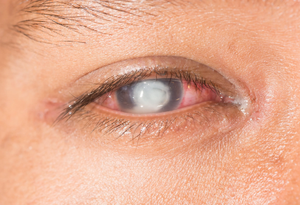
Corneal ulceration (eye ulcer) is an ophthalmic emergency involving epithelial layer disruption and progressive destruction of the underlying corneal stroma. It is an open sore or defect on the cornea, the transparent front surface of the eye. It is usually associated with infection and inflammation, although trauma, contact-lens misuse, severe dry eye, eyelid disorders, and nutritional deficiencies can also contribute. A corneal ulcer may progress rapidly and cause corneal scarring, reduced vision, corneal perforation, or permanent loss of sight if treatment is delayed. The condition requires urgent examination by an ophthalmologist. Diagnosis is based on clinical history, visual-acuity testing, slit-lamp examination, and fluorescein staining. Corneal scraping and laboratory testing may be required to identify the causative organism. Modern treatment centres on intensive topical antimicrobials and surgical repair, while Ayurveda views it as Kshata Shukra or Vrana Shukra—a severe Vata-Pitta and Rakta imbalance—offering complementary cooling and tissue-healing modalities.
Introduction
The cornea is the clear, dome-shaped tissue covering the front of the eye. It allows light to enter the eye and plays an important role in focusing images onto the retina. The cornea contains several layers, including the epithelium, Bowman’s layer, stroma, Descemet’s membrane, and endothelium. The outer epithelial layer acts as a protective barrier against microorganisms, dust, chemicals, and physical injury. When this protective layer is damaged, microorganisms can enter the corneal tissue and produce inflammation. The resulting open lesion is called a corneal ulcer or ulcerative keratitis. As a primary cause of preventable blindness worldwide, corneal ulceration progresses rapidly. A delay of 24 to 48 hours can allow destructive enzymes to melt stromal collagen, leaving behind permanent scars or resulting in structural collapse of the eyeball. It may appear as a white, grey, or yellowish spot on the cornea and is often accompanied by eye pain, redness, watering, photophobia, and blurred vision. A corneal ulcer is considered an ophthalmic emergency because infection can spread into the deeper corneal layers within a short period. Severe disease can lead to corneal thinning, perforation, infection inside the eye, and permanent visual impairment. Early diagnosis and appropriate treatment are essential for protecting vision.
Causes
Bacterial Infection
Bacteria are a common cause of corneal ulcers. Organisms such as Staphylococcus, Streptococcus, and Pseudomonas may enter the cornea through a damaged epithelial surface. Bacterial ulcers may progress quickly, particularly when the infection is caused by aggressive organisms. Contact-lens users are at increased risk when lenses are worn overnight, cleaned improperly, exposed to tap water, or used beyond the recommended replacement period. Poor hygiene can allow bacteria to multiply on the lens or lens case.
Fungal Infection
Fungal corneal ulcers may develop after injury from soil, leaves, wood, agricultural material, or other plant matter. Common fungal organisms include Fusarium, Aspergillus, and Candida species. Fungal ulcers may progress slowly and can be difficult to distinguish from other types of corneal infection. They may produce irregular infiltrates, satellite lesions, or deep stromal involvement.
Viral Infection
Herpes simplex virus can affect the cornea and produce epithelial or stromal keratitis. A typical herpes-related lesion is a branching ulcer called a dendritic ulcer. Recurrent infection may reduce corneal sensation and increase the risk of repeated injury and scarring. Herpes zoster, the virus responsible for shingles, may also affect the eye and cause corneal inflammation or ulceration.
Acanthamoeba Infection
Acanthamoeba is a microscopic organism found in soil and water. It can cause a serious corneal infection, especially in contact-lens users who swim, shower, or wash lenses with contaminated water. Acanthamoeba keratitis may cause intense eye pain, sometimes more severe than the visible eye findings suggest. Diagnosis may be delayed because the condition can resemble bacterial, fungal, or viral keratitis.
Trauma and Foreign Bodies
A scratch caused by a fingernail, plant, dust particle, metal fragment, or other foreign body can damage the corneal epithelium. If bacteria or fungi enter the injured area, the abrasion may develop into an ulcer. Chemical injuries can also damage the cornea and create an opportunity for secondary infection.
Dry Eye and Eyelid Disorders
Severe dry eye reduces the protective tear film that keeps the cornea moist and provides antimicrobial protection. Incomplete eyelid closure, inward-turning eyelashes, chronic eyelid inflammation, and eyelid abnormalities can also cause repeated surface damage.
Other Risk Factors
Other conditions associated with corneal-ulcer development include:
- Diabetes mellitus
- Vitamin A deficiency
- Poor nutritional status
- Reduced immunity
- Previous eye surgery
- Chronic ocular-surface disease
- Long-term or unsupervised steroid eye-drop use
- Poor contact-lens hygiene
- Reduced corneal sensation
- Delayed treatment of eye injury
Pathophysiology
The pathology begins when a breach in the epithelium allows pathogens to attach to the stroma. Microorganisms release exotoxins and proteolytic enzymes (collagenases and elastases) that break down the organised collagen matrix. In response, the host immune system sends polymorphonuclear neutrophils (PMNs) from limbal blood vessels into the avascular stroma. While trying to clear the infection, these neutrophils release reactive oxygen species and matrix metalloproteinases (MMPs). This intense immune reaction causes collateral tissue destruction, known as “stromal melting.” If unchecked, necrosis deepens toward Descemet’s membrane, creating a bulging descemetocele that can rupture into a full corneal perforation.
Signs and Symptoms
The symptoms of a corneal ulcer depend on its cause, depth, location, and rate of progression. Common symptoms include:
- Severe Pain & Foreign Body Sensation: Driven by dense sensory nerve endings in the cornea.
- Marked Redness & Ciliary Flush: Engorged blood vessels encircling the iris junction.
- Intense Photophobia & Epiphora: Hypersensitivity to light accompanied by excessive tearing or mucopurulent discharge.
- Reduced Visual Acuity: Blurring caused by corneal oedema or opaque infiltrates in the visual axis.
- Corneal Infiltrate: A visible grayish-white focal opacity on the clear corneal surface.
- Hypopyon: A layer of pus (inflammatory white blood cells) settling at the bottom of the anterior chamber.
Diagnosis
Medical History
The ophthalmologist will ask about the duration of symptoms, contact-lens use, recent trauma, exposure to soil or plant material, previous herpes infection, eye surgery, medication use, and underlying diseases.
Visual Acuity Testing
Visual acuity is measured in both eyes. This establishes the degree of visual disturbance and provides a baseline for monitoring improvement or deterioration.
Slit-Lamp Examination
A slit lamp provides a magnified view of the cornea, conjunctiva, anterior chamber, and eyelids. It helps determine the size, depth, location, and appearance of the ulcer.
Fluorescein Staining
Fluorescein is a special dye that highlights defects in the corneal epithelium. Under blue light, the damaged area appears green, helping the doctor identify the ulcer and assess its margins.
Corneal Scraping
Corneal scraping may be performed when the ulcer is large, central, deep, atypical, recurrent, or unresponsive to initial treatment. The sample may be examined under a microscope and sent for culture to identify bacteria, fungi, or other organisms.
Additional Evaluation
The doctor may assess corneal sensation, tear production, eyelid closure, intraocular inflammation, and the presence of diabetes or immune-system disorders. Additional testing depends on the suspected cause.
Modern Treatment
The choice of medicine depends on the suspected organism, severity, location, and depth of the ulcer. The general treatment protocol is as follows:
- Topical Antimicrobials: Frequent antibiotic drops; antifungals or antivirals when indicated.
- Cycloplegics: Atropine or cyclopentolate relieve pain and prevent adhesions.
- Surgery: AMT supports healing, CXL strengthens and sterilises the cornea, and therapeutic keratoplasty is used for severe melting or perforation
Ayurveda Insight
In Ayurvedic classics (Shalakya Tantra), corneal disorders affect the Krishna Mandala (dark circular region of the eye). A corneal ulcer corresponds to Kshata Shukra (ulceration due to injury) or Vrana Shukra (ulcerative corneal opacity). The pathology stems from an acute imbalance of Vata (causing sharp pain, dryness, and tissue breakdown) and Pitta (driving inflammation, burning, redness, and suppuration), which vitiates Rakta (blood) and Mamsa (muscle) tissues. Accumulating metabolic waste (Ama) blocks the ocular channels (Srotas), trapping heat and toxins within the eye.
Complementary Ayurvedic care focuses on soothing heat, balancing Pitta, and encouraging tissue regeneration (Vrana Ropana):
- Seka & Aschyotana: Washing the closed eye or applying drops made from cooling herbal decoctions such as Triphala Kwatha or Yashtimadhu (Liquorice) to calm acute irritation.
- Tarpana: During the post-acute healing phase, pooling warm medicated ghee (Maha Triphala Ghrita) over the eye nourishes the stroma and aids tissue repair.
Herbal Remedies for Eye Ulcer by Planet Ayurveda
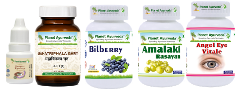
Planet Ayurveda offers a wide range of Ayurvedic medicines and 100% herbal supplements. The products are prepared using carefully selected herbs and traditional Ayurvedic formulations. Each medicine is developed to support specific health needs naturally. The formulations focus on promoting balance, strength, and overall wellness. Planet Ayurveda combines classical Ayurvedic knowledge with quality manufacturing practices. The product range includes herbal capsules, tablets, powders, syrups, oils, and other preparations. These medicines are made to support healthy digestion, immunity, metabolism, and general well-being. Planet Ayurveda aims to make reliable Ayurvedic healthcare accessible and affordable worldwide. In this article, we will discuss some Ayurveda formulations by Planet Ayurveda for the management of Eye Ulcers. Let’s Start!
- Angel Eye Vitale
- Bilberry Capsule
- Amalaki Rasayan
- Mahatriphala Ghrit
- Jasnoor Eye Drops
Care Pack of Eye Ulcer

Product Description
Angel Eye Vitale
Angel Eye Vitale contains Amla (Emblica officinalis), Gotukola (Centella asiatica), and Bhringraj (Eclipta alba), which are traditionally used to support eye health. Amla provides antioxidant support and helps protect ocular tissues from oxidative stress. Gotukola supports collagen formation and may promote healthy tissue repair. Bhringraj is traditionally valued for maintaining clear vision and nourishing the eyes. Together, these herbs may support corneal strength, tissue nourishment, and comfort. Their combined properties may also help maintain healthy inflammatory balance and support recovery in eye-related conditions.
Dosage: 1 Capsule twice daily after a meal with plain water.
Bilberry Capsule
Bilberry Capsule contains Vaccinium myrtillus, a rich source of anthocyanins and other antioxidant compounds that support healthy ocular tissues. These compounds help protect eye cells from oxidative stress and support normal microcirculation around the eyes. Bilberry may also support retinal and vascular health, which contributes to overall visual function. Its antioxidant properties can help maintain healthy inflammatory balance and protect delicate ocular structures. By supporting tissue nourishment and circulation, it may be useful as part of general eye-health care.
Dosage: 1 Capsule twice daily after a meal with plain water.
Amalaki Rasayan
Amalaki Rasayan contains Amla (Emblica officinalis), which is traditionally valued for its cooling, rejuvenating, and antioxidant properties. Its rich vitamin C and polyphenol content may help protect ocular tissues from oxidative stress. Amla supports healthy collagen formation, an important component of the corneal structure. It may also promote tissue nourishment and maintain the natural moisture balance of the eyes. Its Pitta-pacifying properties are traditionally associated with reducing burning, irritation, and heat-related eye discomfort. By supporting ocular strength and healthy tissue repair, Amalaki Rasayan may be beneficial as part of supportive care for eye ulcers.
Dosage: 1 Capsule twice daily after a meal with plain water.
Mahatriphala Ghrit
Mahatriphala Ghrit contains Triphala (Emblica officinalis, Terminalia chebula, Terminalia bellirica), Bhringraj (Eclipta alba), Vasa (Adhatoda vasica), Shatavari (Asparagus racemosus), Guduchi (Tinospora cordifolia), Amla (Emblica officinalis), Yashtimadhu (Glycyrrhiza glabra), Nilotpala (Nymphaea stellata), Pippali (Piper longum), and other supportive herbs processed in ghrita. Triphala and Amla provide antioxidant support, while Bhringraj, Vasa, and Guduchi help maintain healthy inflammatory balance. Ghrita acts as a nourishing base and is traditionally considered beneficial for Vata-Pitta-related eye disorders. Yashtimadhu and Nilotpala may help soothe irritation and support ocular comfort. Together, these ingredients may promote eye-tissue nourishment, support corneal strength, and assist the natural healing environment in eye-related conditions.
Dosage: 1 tsp twice daily after a meal with warm water.
Jasnoor Eye Drops
Jasnoor Eye Drops contain Neem (Azadirachta indica), Shobhanjana (Moringa oleifera), Bhringraja (Eclipta alba), Punarnava (Boerhavia diffusa), Nirgundi (Vitex negundo), and Tulsi (Ocimum tenuiflorum), along with other herbal ingredients. Neem provides cleansing and antimicrobial support for maintaining a healthy ocular surface. Shobhanjana is traditionally valued for its nourishing and antioxidant properties. Bhringraja supports eye strength and tissue nourishment. Punarnava helps maintain healthy fluid balance and may support comfort around the eyes. Nirgundi and Tulsi provide soothing and anti-inflammatory support for irritated ocular tissues. Together, these herbs may help maintain corneal moisture, cleanliness, and a healthy healing environment. The formulation may therefore support comfort and tissue recovery in eye-related conditions such as corneal ulcers.
Dosage: Use 1–2 drops in each eye, three to four times a day, or follow your Ayurvedic doctor’s instructions.
Prevention
Preventive measures include:
- Washing hands before touching the eyes or contact lenses
- Following proper contact-lens cleaning instructions
- Replacing lenses and lens cases as recommended
- Avoiding overnight lens wear unless specifically advised
- Never washing lenses with tap water
- Avoiding swimming or showering while wearing lenses
- Using protective eyewear during hazardous work
- Seeking immediate care after a corneal injury
- Avoiding unsupervised steroid eye drops
- Managing diabetes and chronic dry eye
- Treating eyelid inflammation and abnormal eyelashes
- Avoiding non-sterile substances in the eyes
Conclusion
A corneal ulcer is a potentially vision-threatening condition involving a defect in the cornea and associated inflammation. Bacterial, fungal, viral, and parasitic infections are important causes, while trauma, contact-lens misuse, dry eye, eyelid disorders, and systemic disease increase susceptibility. Pain, redness, watering, photophobia, blurred vision, and a white corneal spot are important warning signs. Diagnosis requires urgent ophthalmic examination, slit-lamp evaluation, and fluorescein staining. Corneal scraping and laboratory testing may be needed to identify the causative organism. Modern treatment includes intensive antimicrobial or antiviral therapy, management of contributing eye-surface disorders, close monitoring, and surgery in advanced cases. Ayurveda offers a traditional perspective based on Dosha balance, Rakta Dhatu (Blood tissue), and Netra (Eye) health. Early diagnosis and appropriate care are essential to prevent corneal scarring, perforation, and permanent loss of vision.
